
Palpitations
Palpitations are a reported symptom and a diagnosis, and describe one’s awareness of the myocardial contraction within the chest, with sensations such as:
- Pounding
- Quick beats
- Skipped beats
They do not necessarily indicate an abnormality of the heart structure, but are often a symptom of an arrhythmia.
Palpitation and its sensation result from the increased blood volume and forced contraction as a result of a prior ectopic beat.
Ectopics appear earlier than a beat would be normally expected on the ECG. They can arise from any region of the heart, but are usually categorised as atrial (PACs), AV junctional (PJCs) or ventricular (PVCs).
*Ectopics can also be called extrasystoles and premature beats.
Depolarisation rate gets slower the further down the heart one travels;
- Fastest in the sinoatrial node
- Slowest in the ventricles
This means that ectopic beats in lower pacemaker sites are usually superseded by those from a superior place.
Patients may not display symptoms despite Ectopic beats occurring regularly.
Atrial arrhythmias are the result of impulses whose origins are outside of the sinoatrial node and stem from three mechanisms:
- Re-entry
- The electrical pathway causes an impulse to travel back into the atrium immediately after it has been depolarised, thus, depolarising it again.
- This will continue as long as the impulse encounters cells that are receptive to it.
- Re-entry can result in atrial, junctional or ventricular beats
- Triggered activity
- The cells depolarise more than once, despite only needing to be activated once
- E.g. Torsade de Pointes
- Triggered activity can result in atrial, junctional or ventricular beats
- Enhanced automaticity
- A pacemaker site other than the SA node takes over the pacemaker function, due to:
- Enhanced automaticity of a non-sinus pacemaker site
- Decreased automaticity of SA node
- Amongst other things, it can be caused by:
- Anxiety
- Fatigue
- Fever
- Narcotics
- Caffeine
- Nicotine
- A pacemaker site other than the SA node takes over the pacemaker function, due to:
Premature Atrial Contractions (PACs)

- Presents with a non-sinus P wave
- Usually followed by a narrow QRS complex
- Can be hidden in T wave, producing a peaked wave
- P wave will display non-sinus morphology and axis
- PACs originating close to AV node produce:
- An inverted P wave
- PR interval ≥ 120ms
- If PAC encounters a refractory AV node, it will fail to be conducted
- Longer RR interval
PAC Terminology:
- Unifocal
- Single focus origin
- All PACs are identical
- Multifocal
- Multiple focus origins
- Different p wave morphologies
- Bigeminy
- Ectopic follows each normal beat
- Trigeminy
- Ectopic follows every third beat
- Quadgeminy
- Ectopic follows every fourth beat
- Couplet
- Two ectopic beats occurring together
- Triplet
- Three ectopic beats occurring together
- Salvo
- Four ectopic beats occurring together
Premature Junctional Contractions (PJCs)

- Pacemaker site junction
- Presents with short PR interval
- <0.12s
- Inverted P waves in:
- II, III, aVF
- P wave can appear around or in QRS complex
Premature Ventricular Contractions (PVCs)
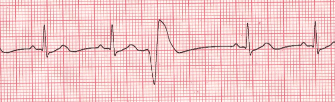
- Ventricular origin, below Bundle of His
- Present with a wide QRS
- >0.12s
- Absent or retrograde P wave
- Right ventricle PVC:
- LBBB morphology
- Left ventricle PVC:
- RBBB morphology

