

Biopotentials are voltages produced by tissues, particularly muscle tissue, during contraction.
In a bit more detail, the concentration of K+ ions is much higher inside the cell compared to the extracellular environment (30-50x) and the Na+ ion concentration is 10x higher on the outside than the intracellular environment. When the membrane is stimulated to about 20mV an action potential occurs and the permeability of the membrane changes, these ions move across the cell membrane and the resulting energies create minute, but measurable potentials on the body’s surface. ECG depends on these electrical signals generated by cardiac muscle to construct a voltage/time graph readable by practitioners.
Due to the nature of the procedure, there are other factors that enable the desired end result to be adequately produced on the trace:
Electrodes
The electrode itself uses an Ag/ AgCl layer to transfer the ion current in the body to the electron current that the monitor uses to create a visible trace.
In practice, there are many different instances in which an ECG will be required, in differing timescales, and on different kinds of patient.
There are various types of electrode and lead clip for these scenarios: smaller electrodes for infants, more adhesive ones for exercise tests and stud clips for more secure lead-electrode connection.
The following must be considered when trying to gain a trace, each can impact the quality of the recording.
- Adhesion to surface
- Electrode gel- electrodes in packets that have been left open can suffer from the gel drying out, which will impact quality.
- Size
- Use-by
- Placement
- Conductor
- Application technique
Skin Preparation
Inadequately prepared skin can have a detrimental effect on the quality of the ECG recording. Errors in amplitudes are common, as are a failure to obtain recordings from leads due to the adhesion being so poor that the electrodes are displaced from the skin surface. The SCST guidelines are as follows:
- Remove excess hair from the electrode sites to ensure maximal contact
- Clean the electrode sites with mild soap and water or a non- alcoholic wipe (alcohol wipes can dehydrate the skin, impeding electrical flow)
- Dry each site vigorously to promote capillary blood flow
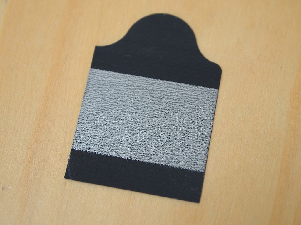
- Light exfoliation of the area using abrasive tape
- Removing part of the stratum corneum allows signals to travel to the electrode
- Abrasion on the stratum granulosum reduces motion artefact

