

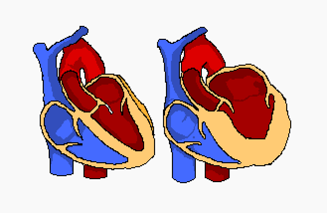
Hypertrophy is defined as the enlargement of an organ or tissue due to the increased size of its cells.
Cardiac hypertrophy then, denotes the increased mass of the heart’s constituent chamber walls. This enlargement is due to an overload, in response to which the chamber in question dilates so as to handle the increased volume of blood within it.
An ECG can pick up the physiological changes in the corresponding area of the heart suffering from hypertrophy, so knowing what area of the heart each section of the PQRST wave corresponds to can help in determining the variety of enlargement.
- Left Atrial Enlargement – LAE
- Increased volume/ muscle mass
- Walls thicken
- Can be caused by:
- Mitral stenosis/ aortic valve disease
- Cardiomyopathy
- Atrial fibrillation (LAE is also often a precursor to AF)
- Hypertension
- LAE on ECG

- Lead II:
- P mitrale
- >0.04s between peaks.
- >0.1s total
- P wave is the sum of atrial depolarisation, so a visible difference is depolarisation speeds in lead II shows LAE
- Lead V1
- Biphasic P wave, terminal negative portion:
- >1mV deep
- >0.04s
- Biphasic P wave, terminal negative portion:
- Right Atrial Hypertrophy – RAH
- Can be caused by:
- Tricuspid stenosis/ pulmonary valve disease
- Chronic lung disease
- Pulmonary hypertension, primary
- Can be caused by:
- RAH on ECG

- P pulmonale
- Inferior leads:
- >2.5mV
- V1, V2
- >1.5mV
- P pulmonale displays as a tall, peaked wave and can often be seen throughout whole trace
- Inferior leads:
- Left Ventricular Hypertrophy
- Can be caused by:
- Aortic valve disease
- Aortic coarctation
- Hypertension
- LVH on ECG
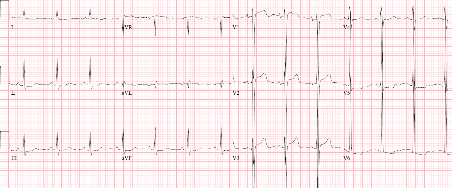
- Dx can be made using a number of formulae, but we will look at the Sokolov-Lyon and Cornell criteria:
- Sokolov-Lyon
- V1 S wave depth + V5 or V6 R waveTallest = >35mV
- LVH present
- Cornell
- aVL R wave + V3 S wave =
- Males >28mV
- Females >20mV
- LVH present
- aVL R wave + V3 S wave =
- Sokolov-Lyon
- Right Ventricular Hypertrophy
- Can be caused by:
- Pulmonary hypertension
- Pulmonary valve stenosis
- Congenital heart disease
- Can be caused by:
- RVH on ECG

- Dx can be made with the following criteria:
- RAD = >+110°
- V1 R waveTallest = >7mV deep
- QRS = <0.12s
- RAH can support Dx
Ref:
Luthra, A. (2007) ECG Made Easy. Third edition. Tunbridge Wells: Anshan
Houghton, A. Gary, D.(1997) Making Sense of the ECG Fifth edition. London: Arnold


One thought on “Hypertrophy”