Tetralogy of Fallot

Recently, in a Holter clinic, I dealt with an 8 year old patient who was on the road to recovery after a diagnosis of congenital defect, Tetralogy of Fallot. As a result, I got hold of the most interesting ECG I have recorded to date.

Background

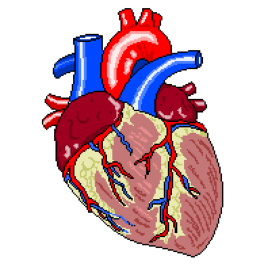
ToF is a rare congential defect affecting the heart, that results in an insufficiency of oxygenated blood leaving the heart through the systemic circulation. Thus, it is considered a cyanotic disorder.

The disorder affects roughly 5 in 10,000 infants, and has an equal gender distribution.

Generally, four pathologies comprise ToF. Whilst all four are not always present, three can consistently be found. ToF is a progressive disorder, in that each pathology gives rise to the others.

The four principal defects are:

RVH

PVSTEN
L-R: Normal and stenotic PV


  • Pulmonary Stenosis
VSD
VSD supplying mixed blood via OvA


  • Ventricular Septal Defect
    • Hole in septum, due to malformation, causing oxygenated and deoxygenated blood to mix within cardiac structure
  • Overriding Aorta
    • Aorta is placed over VSD, transporting blood with low O2 content to wider systemic circulation

Cyanotic episodes require immediate correction, before surgical intervention.

  • High flow O2 administration
  • Physical positioning
    • Knees to chest
    • Parent cradling the child will illicit this effect naturally
  • NaCl fluid bolus
  • Vasopressor therapy
    • Increases systemic vascular resistance, shunting blood through pulmonary system.
  • Continuous ECG and SpO2 monitoring

Surgical intervention usually repairs the VSD and addresses pulmonary pathology, often at the same time.

Prognosis for ToF patients is generally very good.

  • Overall outcome improved since surgical treatment has improved
    • Survival of surgery is currently 95-99%
  • 36 year post-surgical survival is currently 96%
  • Patients who undergo surgical treatment are at greater lifelong risk of ventricular arrhythmia
  • Complications can arise as a result of a transannular patch repair, specifically;
    • RV dysfunction
    • Heart block (risk of HB has dropped to around 1%, in recent studies)
    • Heart failure
    • Recurrent or residual VSD

Hx:

  • 8 y/o
  • Previous diagnosis of ToF
    • VSD
    • PV Stenosis
    • Mild RVH
  • Treatment:
  • Transannular patch repair
  • PV Replacement

Medication:

  • Daily:
    • Atenolol
    • Aspirin

This patient was having a 24hr Holter recording to assess cardiac recovery after their most recent procedure; the PV replacement. Physical examination showed a RVOT murmur, whilst echocadiography displayed a mild RVH and PV regurgitation. Left heart functionality has been classed as excellent.

Previous ambulatory study has shown no arrhythmic action, save for that considered normal in a child of this age. No previous ECG recordings were available.

Upon monitor removal, a 12-Lead ECG was performed, the resulting trace was as follows:

ToFECG (2).png

  • Sinus rhythm with BBB morphology
  • Sokolow-Lyon value of 36mV for RVH
  • QRS & ST segment abnormalities in all leads

Ambulatory analysis relating to the most recent study did not differ greatly from previous monitoring, showing occasional sinus arrhythmia and bradycardia, five non-conducted P waves were found, and two of these gave rise to periods of sinus bradycardia. All other instances were gradual onset/offset.

Nocturnal bradycardia reached rates as low as 34bpm.

What does everyone think of this ECG and brief ambulatory report? Let us know by leaving a comment below!

Heart


Are Athletes At Greater Risk Of Pacing In Later Life?

If so, what is the cause?

The Athletic Heart Syndrome isn’t indicative of any pathology in athletes, and although it is theorised that the changes the heart undergoes as a result of training, there exists no evidence of long-term effects. The athletic heart often has a resting rate much slower than that of an individual of a less active nature. This is not uncommon in physical athletes, as it has been reported that Sir Chris Hoy has a resting HR of 30bpm and fellow cyclist Miguel Indurain one of just 28..!

The cause of this is a very active vagal tone, resulting in bradycardia. As I’m certain many of you are aware, this is a condition that would almost certainly (correct me if I’m wrong) require pacemaker intervention in elderly patients, but in the case of athletes, this bradycardia is due to an increased stroke volume which means the required workload of the heart is decreased. All well and good whilst one is in training, but what if this lower HR did not ‘reset’ to within the normal parameters once training had ceased? I don’t think I’m incorrect in assuming that this would lead to the same treatment a non-athlete, former or otherwise, would receive anyway, regardless of any prior level of fitness.

There is in fact a 2007 study by Baldesberger et al, that suggests this is indeed the case.

Published in the European Heart Journal and found in full here: http://eurheartj.oxfordjournals.org/content/29/1/71 it is shown that there is a statistically significant increase of sinus node disease in the tested former cyclists when compared to the control group, in this case golfers.

Interestingly, I have stumbled across a British Heart Foundation- funded study run in part by the University of Manchester, that they feel suggests the increased presence of arrhythmias in athletes is due to molecular changes as oppose to increased activity in the autonomic nervous system.

The study in rodents showed a decrease in HCN4, a protein found in the mammalian SA node. In humans, a mutation in the HCN4 gene is sometimes found in patients exhibiting sick sinus syndrome and in those who display bradycardia, so the teams behind this study believe that if they can replicate the rodent’s results in humans, it will help us understand arrhythmias that endurance athletes often suffer in later life.

The published study can be found here: http://www.nature.com/ncomms/2014/140513/ncomms4775/full/ncomms4775.html

I’ll answer my second question, “if so, what is the cause?” with an obligatory “je ne sais pas”, but it’s clear that we are edging ever- closer to an answer. Of course, whether that answer is due to molecular changes, or nervous ones remains to be seen.

Either way, it is stated by the team at the University of Manchester that although endurance training can have harmful effects on the heart, these effects are more than outweighed by the benefits.

As an added bonus, here is a short video by Sarah Pratt showing some common differences in an athlete’s ECG (in this case the featured athlete is the NHL’s Tobi Rieder *!*) compared with that of the rest of us. Enjoy!

As ever, if I’ve missed anything, or am just plain wrong about any part of this piece, sound off in the comments below and I’ll do my best to rectify this.

Thanks!

Screenshot (34)

Have You Ever Tested A Robot?

I haven’t. That part comes in a few weeks.

I have, however, BEEN the robot in question, as today, I provided the voice and cardiac controls in my university’s simulation suite.
My peers performed ECGs on a rather frightening, dead-eyed humanoid that was, unbeknownst to them and in ME
conjunction with my voice, being used as a conduit for a scenario pertinent to our learning. That’s me on the right, there, next to my control station (a closer view makes up the header for this post) which allowed me to alter heart rate, breathing rate, create a whole host of arrhythmias and not only see my colleagues, but speak to and hear them as well.

I was a patient named Christopher Smith who had been admitted to A&E. That was all the information that had been supplied, barring my NHS number and date of birth. It was the job of my fellow students to check three patient identifiers, get a brief idea of what was wrong with me and to perform an ECG accordingly, with a brief assessment of the adjustments needed and that of the trace itself.

It was made clear both before and after the session, that it was ok to make mistakes and that this was predominantly what the session was for. It’s extremely unnerving, having a conversation with an expressionless robot that can visibly and audibly breathe, so it was nice to be reassured that the pressure wasn’t as high as it could have been.

Everything going to plan, it would emerge that my chest pain was a result of atrial fibrillation and a heart rate of a mere 32-35bpm. It was also an assessment of how quickly we prioritised the test itself. Due to the presenting chest pains, attaching the limb leads first, so as to gain a visible rhythm strip before a full 12-lead was the correct response, then adjusting the paper speed on the trace itself so as to provide an useable ECG was the next desired step. All the while, I was talking to the student practitioner, asking questions about the test and about the situation in order to see how they reacted and whether they felt comfortable keeping me, as a patient, calm at the same time as carrying out the test with the required level of haste.

These sessions were filmed and then followed a group feedback discussion. The group seemed pleased with the outcome, overall. The comments made were mostly of a positive nature, and the few criticisms there were from myself, my peers and our lecturer, were minor and constructive. This has most certainly been my most enjoyable session to date, and one I did not mind getting up at 4:30am to help set up, so needless to say, I’m very much looking forward to the next one.

I will add that the first half of the session used me as a living mannequin. The reasons that I didn’t comment on this until now are twofold;

  1. It was effectively the same as what I have written about, only without the technology
  2. Seeing my naked torso on film reminded me that I’m still carrying holiday weight. This wouldn’t be a problem, were it not the weight from four holidays.

Thanks!

Screenshot (34)