Healthcare Science Week 2018 – #1

It’s been a little while since I’ve posted to the blog, but Healthcare Science week seems like as good a time as any to do pick it up again. I’ll try my best to upload here or to Twitter, each day, be it information on what might be happening up and down the country, or things going on within my own Trust. I’ve also roped some colleagues into answering a couple of questions, too.

HCS week is an annual event celebrating all 85 specialisms within the discipline. As the oft-overlooked scientists within the NHS, it falls to us to put ourselves out there, and this week is the time to do it! Biomedical Scientists, Physiologists (there are a few different types of this particular scientist), Audiologists, Vascular Scientists, and many, many more are showcasing what they do for patients they treat in the NHS, and using the hashtags #HCSWeek18, and #HCSWeek2018, they’re getting it to the masses. We need to inspire the next generation of scientists to join our ranks, so HCSWeek gives us the perfect opportunity to show students and potential scientists exactly how far-reaching their options are.

Today, I spoke to my colleague Oli, who like me, is a newly-qualified Physiologist, about why he opted to undertake a career as a Physiological Scientist, and what he plans on doing, moving forward.

20180313_164931
Clinical Physiologist, Oli. Here, he is setting up an exercise tolerance test in an attempt to induce ECG changes associated with angina, and ischemia

What attracted you to this particular scientific career in the first place?

It’s as simple as being able to help people, really. Clinical Physiology allows for patient-facing tests to be performed, so I felt like it would let me make an immediate difference.

Now that you’re qualified, and are well-versed in the basics of the job, are there aspects that you appreciate more from a post-graduate perspective?

Definitely. Every day presents a different challenge, because every patient is different. In the RUH, we have a wide array of clinical skills we have to learn, and as a result, I find that I’m adding to my knowledge every day.

Where do you see your career heading, moving forward?

I’m hoping to specialise in Cardiac Imaging, and have applied to the STP programme to help with that speciality. There are a wealth of options, even after that, so whilst I’m not entirely sure at this stage, I may look at the Higher Specialist Training after that!

Heart

AF Association Pulse Check Event

Myself and some of my colleagues recently relinquished a Saturday off, and braved the icy cold (read: British) weather to try to meet with the public, and raise awareness of atrial fibrillation through a project spearheaded by the AF Association.

I didn’t organise the event, but was kindly invited along, and jumped at the chance to help some of my amazing Cardiology workmates by checking pulses, and recording rhythm strips using the AliveCor mobile ECG monitor (which I have previously reviewed here).

AF is an atrial arrhythmia, wherein the sinus node does not cause appropriate, rhythmic depolarisation as it normally would. Rather, multiple foci activate, facilitating a motion akin to ‘quivering’, which raises the risk of embolism through the inefficient pushing of the blood into the ventricles. It’s an incredibly dangerous problem if left untreated, so it’s vital that it gets detected, and preferably this would happen early.

NormAF

I go into detail about AF, it’s mechanisms and ECG presentation in this study guide, so have a look at that if you want to understand it further.

As you may or may not be aware, atrial fibrillation is, globally, the most common clinically significant cardiac arrhythmia, and it is thought that whilst 1.2 million people (a conservative estimate) in the UK are known sufferers of the sinus node disorder, a 500,000 have it, and live undiagnosed. The estimated cost of AF to the NHS was somewhere in region of £2.2 billion, in 2008, and given that the prevalence of the arrhythmia has increased year on year, this number may well be higher now.

Obviously, this is far from an exhaustive exploration of AF, but hopefully it gives some insight into why it’s so important to detect and treat, and why initiatives such as this one are a good idea.

We set up shop in Frome’s Westaway shopping centre at around 10am, where members of the public who’d read about the event in the local newspaper were already queueing. We four clinical scientists proceeded to advise and check 85 people throughout the day. We had a surprise visit from Cardiologist’s Kitchen, too!


IMG_-7mgkhu
Mary, of Cardiologist’s Kitchen fame, showed up to say hello!


Many people we talked to had little-to-no idea what the condition was, its risks, or how it was treated, so we used literature, ECG examples, and a scale model of a heart, to educate, and taught people how to check their own pulse before performing quick rhythm recordings which we analysed on the spot. More than a few people who attended had known AF, and their questions largely involved their current treatment, and the potential impact AF might have on their life. Most, however, visited so they could get checked over, hopefully putting their mind at rest, and learning something in the process.

We didn’t find any new atrial fibrillation (although we did discover two cases of previously undiscovered AV Block), but of equal importance to arrhythmia discovery, was the community engagement, particularly in a public setting. In clinics it’s easy to fall into a cycle with patients, due to schedules and time pressures, and whilst we all try our hardest to make sure everyone is treated individually, seeing the problem before the person is always possible. Interacting with patients on “their turf” meant the ball was in their court, if you like, and the sheer volume of people who expressed an interest meant it couldn’t have been further from a wasted day.

The feedback we received was overwhelmingly positive, and there was a recurring theme in the gratitude people felt for the healthcare environment coming to them, as oppose to the other way around. Many of those to whom we chatted understand the strain that hospitals and GP surgeries are under, and felt that visiting to be checked for AF, and other such things, would be inappropriate. In many ways, I suppose they’re right, too; regardless of the importance of finding these things, especially as they do not always present with obvious symptoms, healthcare centres, unfortunately, cannot cope with the demand a service such as this would present. To this end, I was glad to have ventured out to participate in this, an outdoor clinic of sorts, and educate the public on what to look for, as well as how they can guage their own heart rhythm and take some more control over their own health. I sincerely hope to do it again soon!

20171128_212730.jpg

The AFA is a fantastic charity, so it’d be great if you were to find out a bit more about them by visiting them here.

I’d like to thank my colleagues for asking me to participate, and generally being fantastic people, those who visited us and asked lots of challenging questions, and the kind souls who bought us ginger ale and flapjacks when the temperature reached what *felt* like sub-zero levels.

The statistics used in this post are taken from the BHF. If you want to take a look for yourself, visit the British Heart Foundation, here.

Heart

Is Screening all Young People for Cardiac Disease Cost Effective?

In Western medicine, especially the U.K. and U.S.A., mandatory screening for cardiac disease in young people doesn’t exist.

The NHS currently offers screening for the following:

  • Newborns (blood, cardiac, hearing tests, and physical examination)
  • Pregnancy (foetal abnormalities, blood tests, and infectious disease)
  • Diabetic eye testing (annual follow-ups for cases confirmed in >12 year olds)
  • Cervical (every 3 years for 26-49 year olds, and every 5 years for 50-64 year olds)
  • Breast cancer (50-70 year olds, 70+ individuals can self-refer)
  • Bowel cancer (55-74 year olds)
  • Abdominal Aortic Aneurism (65 year old men. Over 65s can self-refer)

These tests are designed to aid in patient healthcare and to alleviate the long-term financial burden on the heath service, but as you can see, they’re fairly narrow in their scope. Aside from newborns, the vast majority of the screening programmes cater to individuals in later life, or after index presentation/diagnosis. If one suspects an abnormality, then of course, there are many private options available, and in fact, cardiac screening for precursors to sudden cardiac death (such as hypertrophic cardiomyopathy) is in place for young athletes. Given that the estimated number of young people with the abnormality currently stands at 1 in 500, it has been theorised with this and other cardiac diseases in mind, that mandatory screening for young people could ease the cost on the NHS, enhance patient treatment/safety and the emotional fallout that comes as a result of the morbidity associated with serious cardiac conditions.

The results of a nationwide U.K. screening programme known as Cardiac Risk in the Young imply that mass-screening that also encompasses ECG in addition to the standard physical exam and history increases the likelihood of early diagnosis and actually saves money.

The results are the endpoint of a 4-year process, involving roughly 30,000 young persons who were screened using the above methods, all of which were performed and interpreted using the relevant professionals and governing body guidelines.

8.1% of the 30,000 were deemed to produce an abnormal 12-lead ECG, 3.5% had an abnormal physical exam and/or history, and 0.5% showed abnormality in all three areas. 11.7% underwent echocardiography to confirm or deny the presence of dysfunction, 0.9% were referred for cardiac MRI, 1.7% were moved on to longer-term ECG monitoring, and 1.7% had exercise stress testing.

87 abnormalities associated with SCD syndrome were positively diagnosed at the 2-year follow up mark, accounting to 0.3% of the cohort, and 83% of this number was done so using the ECG alone. Interestingly this 83% was found in individuals who presented as asymptomatic, and would likely go undiagnosed otherwise.

The cost of this screening and subsequent treatment of a positive identification is estimated to be 20% lower than the cost of treatment and screening using current requirements, due to its lowering of false positives from 21.8% to 4.3%. This, in turn, lowers the amount of unnecessary follow ups, so this study may have great implications for young athletes, and non-athletes alike in helping to spot these potentially fatal conditions, many of which can be managed.

More on this story and more at: Cardiac Risk in the Young

Heart


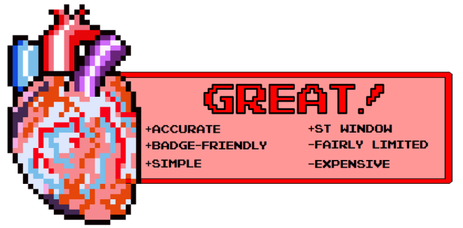
Review: R-CAT ECG Analysis Badge

Price: £9.99/ $8.00

Developer: EKG Concepts (U.S. only. UK Stockist here)

I’m used to reviewing mobile apps and, to a lesser extent, PC and Mac software, so this is something of a new experience for me. I’ve frequently used rate rulers and pocket tools in the past, but the Rapid Cardiac Analysis Tool (R-CAT) is seemingly different to such an extent that I felt it made sense to put it through its paces.

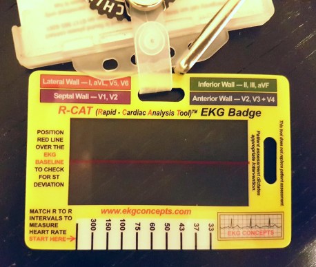
This tool is designed to enable a healthcare professional of any specialism to quickly assess some of the basic criteria of a 3 or 12 lead ECG, such as heart rate, interval and segment duration as well as waveform deviation from the isoelectric baseline.

20161117_000357 1.jpg
I prefer landscape orientation, but the R-CAT accommodates portrait also.

First, the card itself; it’s well crafted, durable, flexible at the same time, is roughly the same size as an I.D. card/driving licence, and its structure and non-embossed build mean it’s easily disinfected, much the same as many other pieces of equipment you would use in a clinical environment. There are two cutaway sections that allow you to attach R-CAT to your lanyard in either a portrait or landscape orientation, and it sits snugly behind the badge holder without getting in the way.

It is, however, too large to slot into the standard NHS card holders which is a minor niggle, but this isn’t too much of a bind as it doesn’t take a huge length of time to remove, and if you have a pull-reel badge holder, it’s no problem whatsoever, as you can use it without removing the card or your lanyard.

The R-CAT focusses specifically on heart rate, segment and interval length, and baseline deviation (if you’re looking for a cardiac axis diagram, then you’ll have to keep looking, I’m afraid). The heart rate measurement works in exactly the same way as a regular rate ruler, with a similar error margin. It doesn’t have as many timing markers as similarly priced full rulers, but I guess it was a necessary concession to make in order to accommodate everything else on its small frame. Whatever the reason, you get a rough to fairly accurate indication of the true R-R interval.

20161117_002249.jpg
R-CAT HR circa 37bpm. The HR given by the equipment and my own calculation was 36bpm


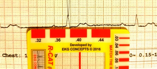
The segment/interval indicators are found on the opposite side of the rate calculator, and span two of the outer edges of the card. The smallest measurement is 0.03s, and the printed values then increase from 0.04s by two, up until the largest at 0.44s. In practice, this system takes a little longer to use than simply measuring with a ruler, but once you’ve used it a couple of times, and get an eye for it, it doesn’t add too many vital seconds to quick analysis.

20161117_003147.jpg
R-CAT measured the PR interval at 0.36s, as did my pencil and paper, and standard rate ruler


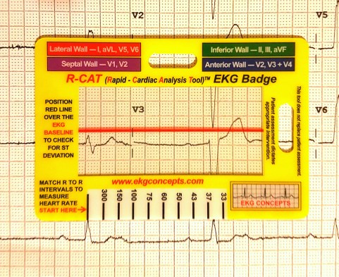
The hook, as it were, that R-CAT introduces, is in the big window through the centre of the card designed to be used in ST segment and Q wave analysis.
Whereas using the edge of some paper, or a ruler can obscure or distort the view of 50% of the waveform under scrutiny, R-CAT uses a thin, red bar in the middle of a clear window, allowing for quick assessment of Q waves and ST segments simultaneously.


20161117_003046
It’s possible to view both positive and negative sections of a waveform at the same time


The window works well, and also functions in the presence of baseline wander, so when such an occurrence is unavoidable, it isn’t a detriment to your analysis.

The company website states it to be more accurate than marking in pencil, as graphite marks can be up to 0.4mm thick, and that is has a greater longevity than callipers, which can loosen with time, and be moved involuntarily. These are excellent points, but with wear and tear printed graphics deteriorate, so I assume that scratches on the measurement indicators could reduce the accuracy of this tool, rendering these comparisons obsolete. Nothing lasts forever, though, and out of the box it works perfectly well.

All in all, I can’t really see this product replacing already-existing products en masse, as this is £10/$8 and the analysis aids already circulating are either slightly cheaper or free, and some provide more functions, but as a learning tool the R-CAT is really effective. During my testing for this review I asked a few cardiology veterans for their immediate impressions, and all shared this opinion. New products in this category are often more of the same; a different colour here, an additional picture there, but R-CAT isn’t one of these products. It shines with its novel portability, and its gimmick; the window. I almost feel bad calling it such, as I feel the word “gimmick” detracts from the product which is, overall, a very cool bit of kit. I won’t say this is an absolutely essential purchase for current professionals who operate outside of an A&E or non-cardiac ward, but it’s certainly worth a purchase. Universities and students however, should take the plunge.

Ah what the heck, it’s unobtrusive and is genuinely helpful when you don’t have a calculator or a rate ruler to hand. If you’ve got a spare £10/$8 kicking about(!), then you should definitely buy one.

rcat-rev


SCST Diploma Day: A Reflection

Myself and OliGS recently sat the SCST Electrocardiography Diploma and Practical Examination, so I thought I’d jot down some of my experiences in the run up, and my retrospective thoughts on the day itself.

If you’re thinking of doing it, or have your PTP finals looming, then read on, as this will give you an idea of what to expect.

I’ll start by saying this: Oli and I have NEVER been so stressed in our entire lives.

This exam was a nightmare for which to prepare; I have extensive experience in taking exams, and it is my view that they’re 50% what you know, and 50% what the examiners want you to tell them. Without having met these examiners or seen a previous paper, it was very difficult to know what to really nail, in the revision stage. The syllabus was long, detailed, and contained what seemed like an entire career’s-worth of things to learn, so we already knew it was going to be a slog, but nothing prepared us for the written paper…

Read these. Lots.

We studied, sometimes sleeplessly, for weeks. Tested each other on rare arrhythmias, read textbooks cover-to-cover (repeatedly), and watched each other’s once sunny outlooks and youthful (ahem) features rapidly wither as the examination date draw closer. It consisted of 20 multiple choice questions, 10 arrhythmia analysis and knowledge questions, and 4 full ECG analysis recordings. Some of these were almost instantly recognisable, but others were brutally difficult to analyse. The MCQs (often the most looked-forward to section of any exam) were equally tough. Those 3 hours lasted a lifetime…

We left the exam battered and bruised, but glad it was over.

But it wasn’t over. It was far from over. As well as the written paper that had almost ruined us, we had the practical exam to do as well.

We had made sure that during our post-ECG placements we still got ourselves in the clinic so as to keep everything fresh, as performing a perfect ECG is not like riding a bicycle.


20161017_221810

The last attempt before test day

It turns out that this was the correct call, as was practicing on one another in the hotel the evening before the exam; the margin for error in the exam is 2mm(!) Anyone who’s had an informal assessment, or had their Direct Observed Practice scrutinised whilst training knows that it’s very easy to second-guess when it comes to electrode placement, and despite having 20 minutes to complete the whole thing, this timeframe becomes devastatingly short once you’re in there. It’s a clinical assessment, so one needs to complete the necessary ID checks, explain the procedure to the patient AND to the examiners (i.e. in two different ways), perform it whist answering questions, and then complete a verbal examination.

Three hours after we had finished the written paper, we were called to attempt our practical exam. There were ashen faces all around. Some were on those still waiting for their number to be called as ours had just been, others had been told that their 2 attempts had been unsuccessful. Neither of us were looking forward to this. Now, given that I’ve already stated that each electrode is allowed to deviate only 2mm from the precise, gold standard location, the internet-purchased electrodes pictured in the above image would be somewhat unfair, right? It seems that the examination board concur, as they provide some rather cool, transparent electrodes complete with crosshairs. They doesn’t make it easier, per se, but they certainly go some way eliminate that lingering trepidation when it comes to deciding you’re happy with your placement and ready for judgement.

temporary_1476735050702.png

Crosshair emblazoned electrodes(!)

I opted for the “all at once” technique: I explained everything to the patient before I started, gained consent, then explained everything I was doing as I went along. Once was put simply to the patient, then once to the examiners, using correct terminology. I paid extra special attention to V1, V2 and V4, as my patient had a particularly wide sternum, so I wanted to be totally sure that I had the sternal border, especially given the electrodes were rather far apart when placed and looked slightly odd to me. After I took a step back and looked at my work, I was incredibly tempted to move the aforementioned electrodes, but either due to fear, fatigue, or a combination of the two, I decided to leave them as they were, opting to go with my initial judgement. After that, I waited.

I’m not entirely sure how the placements are measured, as candidates are asked to leave the room whilst they are checked by two examiners. I heard someone mentioning special rulers, but I didn’t get a look at them (it’s all very cloak and dagger), in any event, you’re called back into the room and, in my case at least, informed of your passing or failing grade. I’m pleased to report that I passed on the first attempt, which as I’m sure you can imagine, was a tremendous relief; I lost my cool somewhat, and expressed my joy rather loudly, as I was informed I wouldn’t have to do it again. Oli soon found me in the waiting area and, grinning wildly, slapped me on the back and hissed “YESSSSS!” before promptly throwing himself into a chair. It was over, and we were victorious.

We didn’t speak too much about it, on the way home, but in the couple of days that have passed since the exam, we both feel a tremendous sense of pride that we actually did it, and did it successfully. If I were to give you all some advice, it would be the following:

  1. Be prepared for anything and everything, including waiting around for a long while
  2. Practice analysing ECGs until you hate them
  3. Practice performing ECGs until you hate them
  4. Go with your gut as much as you can during the practical exam
  5. Bring lunch
  6. Don’t under ANY circumstances, stay at the Ibis Hotel in Birmingham’s Chinatown district (I can’t stress this enough because it backs onto a nightclub that doesn’t stop playing the most bass-heavy music until the wee hours of the morning)

temporary_1469066170176

TSP Mobile: ECG

EDIT: The Android version of TSP Mobile: ECG is available for download, but due to the way in which Google Play operates, I have been unable to offer it for free. The iOS version, when available, will be gratis for the promised 14 days however. Still no word from Apple when that will be, but I have been assured that it is being vetted as I type this, so fingers crossed!

Original article follows:

Well, that TSP mobile app I promised…

I’ve been saying I’d do it for months and, despite remaining fairly quiet with information about starting, I actually have been working on it. So much so, in fact, that the bulk of the development is finished! It’s in final stages of testing, after which it will be available on the Google Play and iOS app stores, where it will be free for the first two weeks of release, so please download it and leave some constructive feedback and a review.

The app features tutorials on ECG analysis, exercise and ambulatory ECG, cardiac flow and cycles, action potentials and useful formulae for trace analysis. Each section is laid out in an easy to follow format, with colourful diagrams and both real and illustrated ECG traces.

Heart rate and QTc calculators are included to aid analysis without leaving the app, and also access to the website blog, so you need never miss an update.

This slideshow requires JavaScript.

I hate advertisements in apps, so in order to keep TSP mobile ad-free, I will charge £1 to download it after these introductory 14 days are over. In an ideal scenario, I would keep it completely free, but it has been, and continues to be, a rather expensive endeavour from both a chronological and economical standpoint especially for my shallow, student pockets, so I hope you understand why I have decided to charge.

Stay tuned to TSP via site, Twitter or email for a release date. It’s very soon!

Heart

Ethical Implications of Switching Off a Pacemaker

On the 24th of September, the BBC reported the story Nina Adamowicz. Nina, a 72 year old lady with an Implantable Pulse Generator (IPG) who, after having the device for almost 20 years, has requested it be switched off.

After suffering a minor infarct, Adamowicz had said that her continued deteriorating health became too much to bear, said she felt like she was waiting in line to be executed, so she requested her device be switched off. She is reported as stating “It isn’t about ‘I want to die’; I’m dying”.

Her case was referred to her local trust’s ethics committee, who, after careful deliberation decided to proceed in line with the wishes of Mrs Adamowicz.

Before passing away on the same night that her device was switched off, Nina Adamowicz stated that she believed she had the right to decide whether or not she wanted the IPG on or off, and stood by her decision.

This case is thought to be the first of its kind in the UK, but Chicago device specialist Dr Westby Fisher professes to doing this exact thing on a dozen separate occasions. Westby considers the ceased action of an implantable device to removing a feeding tube, or switching off a ventilator.

In particular, in a piece for massdevice.com, Fisher tells of a patient who refused dialysis, saying he’d rather let nature take its course. The patient, who also had an IPG, requested that this was switched off, so Westby agreed, and the next day switched off tachyarrhythmia detection on the device. Fisher says that he feels that both he and his patient did the right thing, together.

I for one, am confused as to the ethical pathway involved in coming to both this decision, and that of the ethics committee associated with Nina Adamowicz.. Assisted suicide is complex, but with respect to these scenarios, is defined as the intentional encouragement or assistance to a patient in ending their own life and it is still illegal under the 1961 Suicide Act of UK law . A medic who administers an overdose of muscle relaxants to a patient whose condition is diagnosed as being terminal, even at the behest of that patient, would be punishable by UK law with manslaughter or murder and potentially serve the maximum terms associated with each.

Why then, is hitting the off switch on a pacemaker not considered to be comparable to the example given previously? Patients with implantable devices often have them to combat life-threatening arrhythmias, so in turning them off, this can effectively issue a death sentence to that patient.  I’m not arguing for or against any form of assisted death; I neither understand its intricacies or feel it is my place to denounce or advocate something with which I have had precisely zero experience, I’m simply confused as to why an immediate form of assisted dying is outlawed, and something so similar (on the surface at least), is not. Adamowicz’ clinician has said that other professionals are split in their opinion on his decision, with some feeling it to be “uncomfortably close to euthanasia”.

Is it fair to patients with terminal diagnoses that are forced to travel to countries such as Switzerland, wherein some forms of euthanasia are legal, simply because they do not have an IPG? Does the severity of the condition have any part to play? How similar do individual cases have to be so as to render one illegal and another not so? I have a feeling that this case will spark long debate throughout the medical and legal professions in the UK, and will follow its progress closely.

BBC article

Westby Fisher’s blog

Heart

The Power of Social Media Influence

Like it or loathe it, social media is pretty much inescapable. It’s used by your family, friends, and increasingly by institutions and corporate entities to connect and share ideas, market and promote. Statista puts worldwide social media usage at 2.22 billion people, so it’s no surprise that it has been utilised, and continues to be, to the extent to which we are now accustomed.

It’s been proved that it’s possible to connect with all kinds of people using social platforms, so why should the resource fall solely into the hands of multi-million dollar companies like Coca-Cola and McDonalds, for whom advertising is merely a formality, as oppose to a make-or-break necessity?

Perhaps it needn’t.

Due to the fairly self-regulating nature of some of healthcare’s more specialised areas, the burden falls predominantly on us to showcase innovations and engage with patients, prospective students and fellow professionals. Networking tools like LinkedIn are already being used to connect professionals, even from physiology backgrounds. This platform is relatively self-serving, being a predominantly business to business niche, but according to current statistics it has seen a rise in use to over 60 million views per month in 2016, so is undeniably a great tool to use for quick networking with other like-minded individuals.

Of course, social media can be used to network with everyone, not just our own, so, in the same way that we utilise more than one test to make a diagnosis, we should be using the whole spectrum of tools in this instance, shouldn’t we? Facebook (1.6 billion users worldwide) and Twitter (325 million)  usage polls would suggest that users are logging on for a surprisingly narrow selection of reasons. 68% (Twitter) and 65% (Facebook) of users state that they log on to keep abreast of the latest news relevant to themselves, and 63% and 48% of Twitter and Facebook users respectively, use the platforms to receive information relevant to their personal interests. These present huge, potentially untapped resources for healthcare professionals, that can be used to promote transparency and trust, gain feedback and keep colleagues and patients informed.

I’ve mentioned before, the relatively unknown nature of physiology as a profession, so I think that taking hold of the opportunities available on Twitter, and other forms of social media could be something that could benefit physiological science. One of my favourite online healthcare personalities is Mr Olivier Branford, a plastic surgeon in London. He advocates education as a resource that should be available to all, and public engagement as a high priority. Olivier has over 62.1k followers and uses Twitter to provide news relevant to his specialism, and to wider healthcare in general. I conversed with him about the use of social media as a free platform to provide evidence, studies, inspiration and information to students, prospective students and patients everywhere, and we both agreed that it was the perfect resource to utilise. We aren’t alone, however; Olivier ran a telling informal poll, the results of which I have displayed below, enquiring as to what other users believed was the best way for plastic surgeons to use social media, and I feel that the words “plastic surgeon” can be substituted for any within the health service with a similar outcome. As you can no doubt see; despite the unscientific nature of the evidence, the percentages speak for themselves.

Screenshot_2016-08-01-20-34-19.png

Whilst it would be incorrect to state that healthcare organisations have no presence on social media, they don’t dominate in the same way that more commercial entities do, at least not in the UK. That doesn’t necessarily mean that it is a lost cause, however. Mr Branford has provided a personal touch that corporate entities cannot emulate; his approach of “evidence not opinion” when dealing with healthcare information, is complimented by his willingness to offer an opinion when it’s relevant, on top of the facts. This transparency is refreshing, and, in conjunction with his professional accolades, is surely something that has aided him in gaining  over 62.1k people who want to listen to what he has to say. The cardiac physiology profession is notoriously under-staffed, and whilst the numbers of applicants is on the increase, a quick visit to various college forums shows that the ins and outs of the career are still lost on many students (if you can find a discussion at all). The general career pathways and the salaries seem to be known to these confused individuals, but the actual job is what nobody has much of an idea about. How are we to persuade these potential cardiac scientists to sign up if they don’t know what they’ll be doing for the rest of their professional lives? Asking someone to commit their future to a career and saddle themselves with increasing debt when they don’t really have a great deal of information readily available to them is a far cry from the informed consent we strive to gain from our patients. Taking responsibility, and putting some research into one’s own future is obviously something everyone has to get used to, but I’m sure most people remember how overwhelming that was, so the shortage of new staff members must be more complicated than students simply not looking hard enough. Besides which, it SHOULDN’T be so difficult to find this career..! I’ve got a year to go until I qualify, and I’ve met some truly inspiring people whom, if I wasn’t already on my way, I know could easily convince me to start. We find what we do fascinating, so surely some of these young minds will be just as invested if they have the chance to see it for themselves.

SocMed by age

The Pew Research Centre provides data that places 16-24 year olds as the most avid users of social media (above), and displays a steady growth of users across all age groups year-on-year since 2005, so with a collective effort, it surely wouldn’t be too difficult to a) entice some of these users who are in the middle of their A-Levels, and unsure of which healthcare profession is for them, and b) come together as a profession in a more open and approachable manner to showcase our science and how much of an impact we have on medical diagnostics.

Olivier Branford is a plastic surgeon and associate editor of PRS Global Open journal, and can be found on Twitter under his eponymous handle @OlivierBranford.

Social media statistics obtained from The Pew Research Centre, Statista & Visually

Heart

Review: Epicardio Simulation v1.5 (Full Version)

Download for Windows/OSX:

  • Trial (Free)
  • Paid (£149-£215)
  • 60% discount for full-time students

Developer: Epicardio.Ltd

After my review of the temporary access trial of Epicardio Simulation (which offered a great deal of praise, I might add) I couldn’t wait to have a look at the full version’s features. I still can’t afford it yet even with the 60% discount offered to full-time students, but thankfully, the good people at Epicardio.Ltd allowed me to access the complete package so that I could review it. As I’ve already covered some of the functions of the program, I won’t re-tread old ground, but you can check out what I thought of the trial version here, and consider this a continuation of those original opinions.

So, what functionality is offered by the full version? Let’s go over it now.

The previously-unavailable tutorial section has some marvellous interactive elements; a view of the electrical action and a live ECG accompany the written tutorial pages, allowing the user to view the very thing they’re reading about in real-time. The procedurally generated ECGs are very accurate (I’ve measured them), but if you want to see a genuine patient-obtained trace recording, then one is included with each arrhythmia, too, which really helps with comparisons to the actual recordings one is likely to find in practice.

Almost everything you can think of is covered in some capacity, both on its own, and linked with other, relevant arrhythmias, so you really get a feel for just how interwoven some conduction and rhythm abnormalities can be.

A marvellous inclusion is the level of interactivity within the tutorials; degradation from VT to VF, for instance, is displayed live on the ECG strip and the defibrillator (that I didn’t really have cause to use in the trial version) can be charged, and a shock administered, altering the rhythm strip as it would a real patient.

Screenshot (207).png

The pacing tutorials are easy to use and easy to follow; they walk you through the physics of single and dual chamber, as well as biventricular pacing. In using them to learn the basics of pacing, I can appreciate how effective the arrhythmia sections are and how useful they would have been during the early days of my studies. The interactivity of the aformentioned tutorials remains, too. Placing a pacing wire in different sites allows the user to view live rhythm changes, and sensitivity, HR and pacing rate can be toyed with so as to identify intrinsic rates and pre-pacemaker abnormalities such as 3rd° AV Block on the real-time trace.

Screenshot (212).png

The test area throws generated ECGs at the student, and offers multiple answers from which to choose. Much like any degree-worthy multiple choice test, they range from incredibly easy to downright tricky, but a review section allows you to view the areas that might require further learning before each future run-through. As with the main bulk of the software, measurement calipers are useable during the test, allowing for some precise questions to be given.  Importantly, this software allows and encourages repetition; fundamental to successful learning. It may seem obvious, but I noticed that my understanding of unfamiliar areas increased the more I explored them. What won’t be obvious, is just how quickly this occurred. With the addition of the test function, the user can consolidate what they have learned at their own pace, and not have to exit the program find a different testing app.

Screenshot (217).png

My time with the trial version of Epicardio only threw up a couple of minor issues. Whilst these are still present, they detract from the simulator even less than before, due to the myriad of extra content present in the full release. My only new problem came in the single chamber pacing tutorial, wherein I was instructed to reduce the pacing rate to 45bpm, yet I couldn’t lower it past 50bpm. This made it impossible to view the intrinsic rhythm of the digital patient (the point of the page in question’s existence), but only in this instance. It’s worth pointing out that regular updates exist to iron out glitches such as this, so errors needn’t remain for long.

Screenshot (216)

If, like me, your learing speed is increased by doing, as oppose to just reading or seeing, then you’ll find this tool invaluable. To be able to safely induce life-threatening ventricular rhythm is, understandably, an uncommon occurrence, so a method to facilitate this, and things like it, is always going to be welcome for students. In Epicardio, however, you get so much more than that. Pacing of all types is covered in depth, real and digitally created ECGs, and an effective test facility really do set this above any of the other programs that I’ve used. It’s also incredibly simple to get the hang of, too. The things it does well far outweigh its minor issues, so I can wholeheartedly recommend this program to everyone who wants learn about cardiac arrhythmia and interventions. Whilst the implementation of a 60% student discount brings the price down to the £59-£89 mark, it is still expensive, but you really do get what you pay for.

Screenshot (215).png

Heart

Tetralogy of Fallot

Recently, in a Holter clinic, I dealt with an 8 year old patient who was on the road to recovery after a diagnosis of congenital defect, Tetralogy of Fallot. As a result, I got hold of the most interesting ECG I have recorded to date.

Background

ToF is a rare congential defect affecting the heart, that results in an insufficiency of oxygenated blood leaving the heart through the systemic circulation. Thus, it is considered a cyanotic disorder.

The disorder affects roughly 5 in 10,000 infants, and has an equal gender distribution.

Generally, four pathologies comprise ToF. Whilst all four are not always present, three can consistently be found. ToF is a progressive disorder, in that each pathology gives rise to the others.

The four principal defects are:

RVH

PVSTEN
L-R: Normal and stenotic PV


  • Pulmonary Stenosis
VSD
VSD supplying mixed blood via OvA


  • Ventricular Septal Defect
    • Hole in septum, due to malformation, causing oxygenated and deoxygenated blood to mix within cardiac structure
  • Overriding Aorta
    • Aorta is placed over VSD, transporting blood with low O2 content to wider systemic circulation

Cyanotic episodes require immediate correction, before surgical intervention.

  • High flow O2 administration
  • Physical positioning
    • Knees to chest
    • Parent cradling the child will illicit this effect naturally
  • NaCl fluid bolus
  • Vasopressor therapy
    • Increases systemic vascular resistance, shunting blood through pulmonary system.
  • Continuous ECG and SpO2 monitoring

Surgical intervention usually repairs the VSD and addresses pulmonary pathology, often at the same time.

Prognosis for ToF patients is generally very good.

  • Overall outcome improved since surgical treatment has improved
    • Survival of surgery is currently 95-99%
  • 36 year post-surgical survival is currently 96%
  • Patients who undergo surgical treatment are at greater lifelong risk of ventricular arrhythmia
  • Complications can arise as a result of a transannular patch repair, specifically;
    • RV dysfunction
    • Heart block (risk of HB has dropped to around 1%, in recent studies)
    • Heart failure
    • Recurrent or residual VSD

Hx:

  • 8 y/o
  • Previous diagnosis of ToF
    • VSD
    • PV Stenosis
    • Mild RVH
  • Treatment:
  • Transannular patch repair
  • PV Replacement

Medication:

  • Daily:
    • Atenolol
    • Aspirin

This patient was having a 24hr Holter recording to assess cardiac recovery after their most recent procedure; the PV replacement. Physical examination showed a RVOT murmur, whilst echocadiography displayed a mild RVH and PV regurgitation. Left heart functionality has been classed as excellent.

Previous ambulatory study has shown no arrhythmic action, save for that considered normal in a child of this age. No previous ECG recordings were available.

Upon monitor removal, a 12-Lead ECG was performed, the resulting trace was as follows:

ToFECG (2).png

  • Sinus rhythm with BBB morphology
  • Sokolow-Lyon value of 36mV for RVH
  • QRS & ST segment abnormalities in all leads

Ambulatory analysis relating to the most recent study did not differ greatly from previous monitoring, showing occasional sinus arrhythmia and bradycardia, five non-conducted P waves were found, and two of these gave rise to periods of sinus bradycardia. All other instances were gradual onset/offset.

Nocturnal bradycardia reached rates as low as 34bpm.

What does everyone think of this ECG and brief ambulatory report? Let us know by leaving a comment below!

Heart