Synap: Beta Test Update

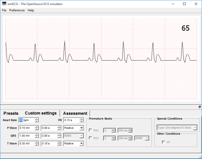
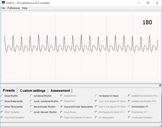
Synap is an upcoming revision tool that is driven by students. The platform enables students to create their own multiple choice questions and upload them, then download those created by others. It’s possible to “follow” other users, as you would someone on Twitter or, incidentally, this site (you can do that in the sidebar of this page…), and take any quiz that they have created. Image upload and basic editing is supported, so quizzes for physiology, such as ECG arrhythmia or echocardiography quizzes are more than possible, and are one of the reasons I decided to get involved with the whole thing. In addition, the app tracks your progress and structures your revision for you, based on your course and modules.

I’ve spent the last week or so beta testing the Synap web platform or, more specifically, I’ve been taking tests and creating basic ECG quizzes to help bug test and check functionality.

The platform, as I’ve mentioned, is currently in closed beta and only present on the web, so without having an app and a larger number of users I cannot comment on it fully, but as it stands, the processes involved in creating a profile and quiz are incredibly simple; adding and annotating images is a cinch, and a complete question only requires the user to add a correct answer and a few wrong ones. Whilst I encountered a few bugs initially, the feedback I provided was swiftly taken on board and the problems were remedied overnight. Taking quizzes is incredibly simple, and all you need to do is click “take quiz” (shockingly), then select your answers and have them marked. You can take these as many times as you like, too, and if the creator has provided any, feedback will be available for each individual question.

My only concern is the reliance on the quiz-maker supplying the correct information. I’ve taken a quiz wherein the correct answer was the only one that was possible to be correct (think “What has tusks and a trunk? 1) Elephant 2)Belephant 3)Your hamster”) yet I was still told my answer was wrong. This is a closed beta, though and that’s what these processes are for. I know it hasn’t escaped the attention of the developers, so we shall see how it is dealt with.

To break all of this down and show you what I know for sure so far, have a look at this (incomplete) features list:

  • MCQs:
    • Image/annotation upload
    • Correct answer & up to 5 incorrect
    • Optional feedback for test-taker
    • Optional link to external learning resource
    • Test result calculation
    • Obtainable achievements
    • Personalised revision quizzes sent to you
  • Community links based on:
    • Course/Discipline
    • Cohort
    • Institution

I’ll add to this list the more familiar I become with the platform.

Omair and James, its creators, and the rest of the Synap team hope that this app will enable students nationwide to help each other and revise together, and it’s a pleasure for me to be involved, even if it’s only in a small way, currently. I’ll continue to post updates as things progress.

For more info, visit @Synap on twitter.

Thanks

Screenshot (39)


Holiday-Only Arrhythmias

During the festive season, its easy to indulge in excess; too many sprouts, an increase in afternoon napping, festive drinks… You know the score. It isn’t all smiles and sunshine, though, as we shall see.

One particular result of all the festive excess relevant to cardiac professionals, has been reported across the globe, but particularly in Entirely Fictitious Primary Care Centres (EFPCCs); Bacardi Branch Blocks, or BacBBs

BacBBs are thought to affect the heart as a whole, but it can be seen that they have a particularly odd effect on the ventricles, and cause an odd, never-seen-in-real-life depolarisation wave on the ECG, that actually defies physics and medical science by going back in time!

Symptom sheets compared with the compiled ambulatory data have shown unanimously that BacBBs are present sporadically within sinus rhythms, but coincide with that one-drink-too-many during a family game of Monopoly (Mr Moneybags isn’t thought to be an underlying cause, so the activity isn’t seen as a risk factor).

Atrial activity stops altogether, presumably because the SA node just forgets what it’s doing, as it’s seen enough crepe paper hats and screwdriver sets fly from crackers to last it a lifetime.

After an episode of BacBB, sinus rhythm resumes, and the patient will return to whatever their festive-norm may be until the next instance.

This phenomenon seems to disappear entirely during the first couple of weeks of January, when normal working hours begin again, hence, I feel that it is triggered by the holidays themselves.

None of this is being researched, or is even disputed, because it is both totally false, and invented entirely by me.

Screenshot (130)

Bacardi Branch Block

  • Common holiday rhythm abnormality only found during the festive season, and even then, only in fictitious settings
  • HR between 80-120bpm
    • Depends entirely on board game leader-board position
  • No P waves
  • Abnormal ventricular action
    • Resembles upturned cocktail glass
  • Is thought to only contribute to familial tolerance levels during prolonged exposure to each other


HAPPY HOLIDAYS FROM EVERYONE AT THE STUDENT PHYSIOLOGIST!!

Screenshot (124)


ECG Difficulties

I never considered just how difficult trace analysis could be. Don’t get me wrong; I knew it would be hard, I just didn’t fully appreciate quite how hard.

During lectures on specific arrhythmias, when ECGs are displayed, they generally contain the abnormalities that make up the subject matter so it doesn’t take long to come to the correct answer, but looking at a trace without any history or prompting as to the condition, is still overwhelming to me. So overwhelming, in fact, that I often feel like I’m falling short of the mark with regards to my learning as a whole. The TSP ECG section is as much for my benefit as it is for you guys, in that I’ve found analysing the ones selected for posting incredibly difficult.
No matter what answer I come to, there’s always the lingering worry that I’ve missed something.

How much is too much, with regards to analysing?

What’s a result of over-analyzing, and what’s accurate?

Textbook traces, whether clinical, or stylised, have been selected as the best possible example of the rhythms under scrutiny, so it stands to reason that they won’t exactly mimic those that will be encounered in the field. In my limited experience, clinical traces contain a great deal of variation and have thus far, rarely resembled anything you’d find in a book.

They have been difficult, yes, but they have also been possible. This will all become easier, with practice (I assume/hope), so I hope you all find the analysis quiz good practice, as it’s certainly proving to be that for me.


Screenshot (124)

Pathological causes of LVH

My last article looked at the assessment of Left Ventricular Hypertrophy; its contextual clinical significance and subsequent electrocardiographic findings, and concluded with possible pathological reasons for the development of LVH of which I wanted to discuss in my next article.

Sadly, due to an onslaught of assignments more intimidating than Xerxes Persian army in the film 300, I haven’t had the time to write any subsequent material.

However, now the assignments are over I have the time to explore these pathological causes of LVH.

Just as a recap, LVH is an increase in the size and proportion of the left ventricular myocardium. Just like any muscle, the more it is permitted to carry out work (contract) the greater it will increase in size (hypertrophy).

This increase in muscular size results from increased recruitment of sarcomeres (basic subunit of muscle cells) as well as extra cellular matrix remodeling (the scaffolding material of tissue). As a result of these anatomical adaptations the ventricle changes in size and proportion. Its normal conoid shape may be altered.

Concentric/Eccentric Hypertrophy

This remodeling will present as either Concentric or Eccentric hypertrophy depending on the underlying cause.

Concentric hypertrophy results from chronic pressure overload commonly associated with chronic hypertension and aortic stenosis. New sarcomeres are added in parallel to existing sarcomeres. Wall thickness greatly increases and persistence over time will significantly reduce chamber radius. The remodeled ventricle has reduced contractility  and compliance leading to diastolic and eventually systolic dysfunction (impaired filling/ejection).

Eccentric hypertrophy often occurs with volume and pressure overload; pathological associations include heart failure; aortic/mitral regurgitation (volume overload) and chronic hypertension (pressure overload). Ventricular remodeling results in increased chamber radius and moderate increases in wall thickness. Chamber dilation occurs as new sarcomeres are added in series to existing sarcomeres.


Figure-4-Concentric-and-eccentric-hypertrophy-can-be-distinguished-by-the-orientation-in

Physiological consequences of LVH

LVH usually develops as a compensatory response to the underlying pathologies mentioned above. Increased arterial pressure (afterload) as a result of chronic hypertension and/or aortic stenosis increases the pressure required of the LV to eject this blood.  Increased LV wall tension compensates via concentric hypertrophy.

Volume overload within the heart (heart failure) is often a resultant of valvular regurgitation and/or systolic dysfunction. Aortic/mitral regurgitation will increase the volume of blood left in the ventricle after systole (End Systolic Volume). During the next systolic cycle the LV has to contract with greater force to eject this increased volume of blood (End Diastolic Volume). Frank Starlings law of the heart states that increased stretch on the myocardial wall (Preload) increases strength of contraction. This pressure/volume overload induces chamber dilation and eccentric hypertrophy.

The hypertrophied LV becomes less compliant reducing its filling and contractile capacities. This culminates in systolic dysfunction. Systolic dysfunction is a significant reduction in cardiac output and will present with symptoms of dizziness, fatigue and shortness of breath. Systolic dysfunction of the LV will also lead to pulmonary congestion due to the back up of pressure generated by increased atrial and pulmonary venous pressures resulting from the increased EDV.

LVH is one of the strongest predictors of cardiac morbidity in hypertensive patients. The degree of hypertrophy correlates with the development of congestive heart failure, angina, arrhythmia, myocardial infarction and cardiac death (Lilly).

Another pathological subcategory I have not eluded to that is also a major contributor to LVH is cardiomyopathies. This is something I will look at in detail in my next article. Thanks for reading 🙂


I’d just like to take the opportunity to thank my good friend and partner in crime Christopher Wild for firstly creating this fantastic physiology based resource and secondly giving me the opportunity to participate in its progression.

3 months since creation and the TSP has already received nearly 1500 hits, recognition and support from numerous universities and academics across the country as well as our professional governing body.

My buddy deserves massive acclamation for this achievement and I know there is much more to come!

Whilst writing this article it has again reminded me how interconnected many pathologies, symptoms and clinical findings can be. About half way through writing I felt as though I’d opened a big can of worms as there are so many different tangents on which you could proceed to discuss. Added to this is the limited knowledge I have as I’m only a second year student! Therefore please don’t take this information as cardiology gospel! I have and always will, use reliable sources of information, but this is my interpretation of such material and I can’t guarantee inclusion of every detail.  Nevertheless, I have personally found writing such articles to be of great benefit; and thus if there are any other physiology students out there that may be interested in writing for TSP we would greatly welcome your support.

Screenshot (39)

Ref:

Lilly, L. S. (ed.) (2010) Pathophysiology of heart disease: A collaborative project of medical students and faculty. Editor, Leonard S. Lilly. 5th edn. Philadelphia, PA: Lippincott Williams and Wilkins.
(Lilly, 2010, pp. 315 – 315)

http://www.cvphysiology.com/Heart%20Failure/HF009.htm

Image from:

https://www.researchgate.net/figure/51745685_fig2_Figure-4-Concentric-and-eccentric-hypertrophy-can-be-distinguished-by-the-orientation-in


Postgraduate Preceptorship

One of the SCST Annual Update sections most relevant to PTP students was based around postgraduate preceptorship. Delivered by Sophie Blackman, of Boston Scientific and the SCST, the talk went through the updates regarding the assessment and accreditation that a newly qualified physiologist can obtain, in conjunction with the council. Sophie stated that she has been quite heavily involved in the production of this framework, and proceeded to outline its intricacies.

The preceptorship programme is relevant to PTP students, because it is aimed specifically at us once we are “let loose”, as it were, into clinical practice. The SCST has received a great deal of feedback from around the country, pertaining to the varying levels at which new healthcare scientists are emerging from their academic study and also feedback from students themselves, on what they feel they need by way of support from the governing body. I for one, can appreciate this; I’m terrified of graduating. I’m confident in my own skills, thus far, and whilst I don’t think that fear will ever go away, I think it’s beneficial for newly qualified HCS to have someone outside of their department that can help them make that transition from student to professional. This nationwide initiative will help to provide this support for the individual, but will also ensure there is an equitable workforce in practice.

The programme itself is based around the individual, and is likely to take anywhere from six months, to two years, based on proficiency. Much like in your current degrees (if you’re a student), the programme features competencies and case based discussions, but in this case, they are undertaken as you perform a job at which you are already doing. Upon “qualification” (this may seem like an odd word to use, given the fact that the practitioner is already qualified, but bear with me…), the student will receive a certificate displaying their confidence in a particular discipline, and that can not only demonstrate a willingness on the practitioner’s part to be the best that they can be, but it will further cement that person’s knowledge and skills base to aid them in their position, thereby helping them, to a point, to leave the student role behind them.

The implication was that if you, as a new HSP, want to make that leap to the STP programme, or follow a different career framework, then this accreditation will assist in identifying your individual fortes, and allow you to perhaps see what pathway you would be best suited to.

The full texts relevant to students are given below:

Preceptorship Framework

Preceptorship guidance for HSPs

For more information, visit the SCST preceptorship page:

new_scst_logo

Screenshot (39)


Review: AliveCor Mobile ECG Monitor (3rd gen)

Download the android app: Free

Purchase the monitor: £75 approx.

Developer: AliveCor, David Albert

Thus far, my reviews have been mostly confined to apps, with the only exception being Windows/Linux software, simECG. This review is quite exciting for me, as it involves a physical monitor as well as a companion app. I picked up the now world-famous AliveCor Mobile ECG Monitor a couple of days ago to road test it, and I’m pleased to say that for patients, it’s fantastic, and for students, it’s just as good.

As far as functionality goes, this app serves as a personal event monitor with a particular focus on atrial fibrillation, and it has a ton of nice features that make it a worthwhile investment for patients regular to cardiac departments.

alivecor-fda-cleared-alivecor-mobile-ecg-heart-rate-monitor-for-400x400-imae6ajchafcryr8

Out of the box, the dual-electrode plate can be attached to the back of your mobile device via an adhesive strip, or kept separate; AliveCor works either way, and if you do attach the monitor and change your phone, you can pick up additional attachment plates for around £6.

Obtaining a trace is a very quick process; it only took me a few seconds to open the app and begin recording, and the trace is saved automatically after 15 seconds, with the limit set at 30. The user is then presented with a series of tick-able boxes such as hand or chest ECG, and a notes section to document any symptoms. These are then stored with the trace.

In this video, you can see that AliveCor jumps straight into recording once fired up.

Heart rate and beat fluctuation are tracked and graphed automatically to allow patients to relate multiple recordings in conjunction with the particular activity being performed during monitor operation.

Screenshot_2015-11-30-09-13-23~2

In addition to this, the app comes with an algorithm that determines the presence of atrial fibrillation and keeps track of how many instances this occurs.

AliveCor offers a great deal of options when it comes to sharing data and to physical useage: once the trace has been recorded, the user can email it, save it as a fully notated PDF and print either from the app or a different program.

Screenshot_2015-11-30-13-16-10~2

Holding the device in your hands, as shown in the app instructions gives you a trace in lead I, and it’s possible to obtain leads II and III by placing the two electrodes in different areas of the body (I have provided these instructions at the bottom of the page). Handily, AliveCor doesn’t just measure biopotentials in the peripherals, but also in the chest. A Lewis lead configuration is possible to view atrial activity with more clarity.

I experienced a minor issue with artefact at the start of recording, but this was almost definitely user error, as AliveCor ‘steadies’ itself pretty quickly if you remain relaxed and support you arms. This learning curve is honestly the only problem I had with the product, and after 10 or so minutes, it wasn’t a problem at all. I don’t want to speak for everyone, but I feel it’s fairly easy to get to grips with, so I doubt that your average patient would have too much trouble with it after a short while.

Traces themselves look very clean and, thanks to the standard calibration and the inclusion of a regular ECG paper grid, various amplitudes, intervals and waveforms can be measured manually. The trace screen also gives the option to invert the recording, and filter enhancement is selectable for each one.

As an event monitor, this device is invaluable. It comes with its own built-in symptom sheet and it’s incredibly quick and easy to record a good quality trace. AliveCor has been given the thumbs up from the FDA and NICE, so it’ll be interesting to see how the SCST view the monitor; I’ve reached out to them, but haven’t heard anything yet. If I do, I shall update accordingly.

I assume that in the U.S. this app allows patients to forgo some of the high cost of continued medical care by way of allowing the trace to be sent directly to a clinician for review. The UK version gives the option to send the trace to a Cardiac Physiologist for £5 and provides the analysis results within 24 hours, allowing the patient to present an official ECG report to their GP, should they need to.

As an added bonus, the AliveCor app has an educational area that features breakdowns of common arrhythmias and cardiac anatomy. The illustrations are aesthetically very pleasing and straightforward. The information contained within it is not as comprehensive as the information you’ll find in your lectures or textbooks, but it isn’t designed for the use of practitioners, so what is there is entirely sufficient.

All in all, AliveCor truly is a technical feat and not only does exactly what it sets out to do, but gives a glimpse of the future of ECG technology. This is an extremely good way for patients to become actively involved in their own heart health, with a relatively small price tag. The app provides a simple, intuitive UI and doesn’t require any Bluetooth connectivity between monitor and phone: it works right out of the box so that any patient can use it with ease. There’s a reason this product has garnered praise around the globe.

I will add that the device’s creator, Dr David Albert, is one of the nicest individuals with whom I have ever had the pleasure of conversing. His instructions for getting the most out of AliveCor for the purposes of this review have been invaluable, and even though he really didn’t have to, he answered every question I asked him, swiftly too. I’d like to thank David for being kind enough to help me get to grips with the product all the way from his residence in Oklahoma. Students need input such as this; it cements that we are valued and encourages learning outside of regular studies.

Screenshot (183)


Screenshot (39)

Positioning data:

Lead I: LH – RH

Lead II: LL (knee) – RH

Lead III: LL – LH

Lewis: Electrode 1 on V1, device angled vertically

Review: simECG: ECG Simulator v1.186

Download for Windows/Linux: Free

Developers: Antonio Cardoso Martins, Paulo Dias Costa, Joao Miguel Marques


I’ve been searching for a half-decent ECG simulator since last year, but hadn’t found one that costs less than “more than I have”, so I was pleasantly surprised to find the rather unnecessarily named simECG: ECG Simulator for free, on Windows and Linux.

simECG offers a number of functions in its current version. The operator can select from a series of common arrhythmias at the click of a button, and observe the associated waveform on the display. Unfortunately, only a handful of options are actually selectable, at present, with the others showing as greyed out, presumably, as with many Open Source programs, until they are finalised by the development team.

simECG screen 2 (2)

The custom settings tab provides the means to alter each area of the trace individually, adjusting heart rate, P wave amplitude/duration and more, and watching the displayed trace change in real time. The program hints at future save/load functions for your altered settings, too, which will be a nice inclusion for educators to make use of.

simECG screen 1

All of the aforementioned are easy to use and clearly marked, even if there aren’t currently all that many of them.

The option exists to change the background between ECG paper and a monitor screen, although the ECG paper skin is purely cosmetic. It would have been nice if the paper option was more in correlation with the amplitudes and durations selectable in the readout options. Greyed out sections of the “preferences” tab hint that calibration will soon be able to be changed by the user, so it would be preferable for beginners and students if these proposed calibration options had a realistic background to use in conjunction with the created trace.

simECG screen 4

I couldn’t find an option to reset the trace at all, even in a greyed out form, and as a result, returning to the default custom settings is something of a chore. Hopefully this is something the developers will consider including in future iterations.

By now you may have noticed the appearance of the waveforms in the above trace. The trace waveform was one of the first things I noticed, as the whole thing doesn’t look right. The P and T waves look malformed, with the latter presenting almost as though the patient was displaying hyperkalaemia despite this being labeled as a normal sinus ECG.

simECG screen 3

The assessment quiz tab gives the user an opportunity to identify 10 rhythms in 60 seconds. It’s fun, sure, but given the odd appearance of the waveforms, it becomes a case of memorising the traces present in this program alone, as they aren’t all applicable to real life.

I’ll be honest, it’s hard to criticise something that the developers admit will “never be finished” due to its Open Source status, but the nature of this website and Open Source in general means it pays to remain objective. In actuality, whilst I have highlighted a few issues, the fact that this tool is ever-evolving and totally free, means I can only commend the development team for their ethos and hard work.

Martins, Costa and Marquez state their belief that education shouldn’t be a corporate tool, or purchasable commodity, rather it should be accessible to all. The more people there are to flag issues, the better an idea the team can have of what functionality to add, what bugs to fix, and what other changes are felt to be necessary by users. Despite being generally incomplete at present, it’s not only one to watch for in the future, but one I’d ask every cardiac physiologist to download and play around with.

Due to this version still being in the 1.n phase, I have high hopes for the future of this software, as it has great potential as a learning tool. With the addition of more options in the preset tab, further wave/interval customisation, and more accurate waveforms in general, simECG could help physiology students consolidate their knowledge without carting loads of textbooks around, making it an essential bit of kit.

Screenshot (184).png


Screenshot (39)