EDIT: The Android version of TSP Mobile: ECG is available for download, but due to the way in which Google Play operates, I have been unable to offer it for free. The iOS version, when available, will be gratis for the promised 14 days however. Still no word from Apple when that will be, but I have been assured that it is being vetted as I type this, so fingers crossed!
Original article follows:
Well, that TSP mobile app I promised…
I’ve been saying I’d do it for months and, despite remaining fairly quiet with information about starting, I actually have been working on it. So much so, in fact, that the bulk of the development is finished! It’s in final stages of testing, after which it will be available on the Google Play and iOS app stores, where it will be free for the first two weeks of release, so please download it and leave some constructive feedback and a review.
The app features tutorials on ECG analysis, exercise and ambulatory ECG, cardiac flow and cycles, action potentials and useful formulae for trace analysis. Each section is laid out in an easy to follow format, with colourful diagrams and both real and illustrated ECG traces.
Heart rate and QTc calculators are included to aid analysis without leaving the app, and also access to the website blog, so you need never miss an update.
This slideshow requires JavaScript.
I hate advertisements in apps, so in order to keep TSP mobile ad-free, I will charge £1 to download it after these introductory 14 days are over. In an ideal scenario, I would keep it completely free, but it has been, and continues to be, a rather expensive endeavour from both a chronological and economical standpoint especially for my shallow, student pockets, so I hope you understand why I have decided to charge.
Stay tuned to TSP via site, Twitter or email for a release date. It’s very soon!
Like it or loathe it, social media is pretty much inescapable. It’s used by your family, friends, and increasingly by institutions and corporate entities to connect and share ideas, market and promote. Statista puts worldwide social media usage at 2.22 billion people, so it’s no surprise that it has been utilised, and continues to be, to the extent to which we are now accustomed.
It’s been proved that it’s possible to connect with all kinds of people using social platforms, so why should the resource fall solely into the hands of multi-million dollar companies like Coca-Cola and McDonalds, for whom advertising is merely a formality, as oppose to a make-or-break necessity?
Perhaps it needn’t.
Due to the fairly self-regulating nature of some of healthcare’s more specialised areas, the burden falls predominantly on us to showcase innovations and engage with patients, prospective students and fellow professionals. Networking tools like LinkedIn are already being used to connect professionals, even from physiology backgrounds. This platform is relatively self-serving, being a predominantly business to business niche, but according to current statistics it has seen a rise in use to over 60 million views per month in 2016, so is undeniably a great tool to use for quick networking with other like-minded individuals.
Of course, social media can be used to network with everyone, not just our own, so, in the same way that we utilise more than one test to make a diagnosis, we should be using the whole spectrum of tools in this instance, shouldn’t we? Facebook (1.6 billion users worldwide) and Twitter (325 million) usage polls would suggest that users are logging on for a surprisingly narrow selection of reasons. 68% (Twitter) and 65% (Facebook) of users state that they log on to keep abreast of the latest news relevant to themselves, and 63% and 48% of Twitter and Facebook users respectively, use the platforms to receive information relevant to their personal interests. These present huge, potentially untapped resources for healthcare professionals, that can be used to promote transparency and trust, gain feedback and keep colleagues and patients informed.
I’ve mentioned before, the relatively unknown nature of physiology as a profession, so I think that taking hold of the opportunities available on Twitter, and other forms of social media could be something that could benefit physiological science. One of my favourite online healthcare personalities is Mr Olivier Branford, a plastic surgeon in London. He advocates education as a resource that should be available to all, and public engagement as a high priority. Olivier has over 62.1k followers and uses Twitter to provide news relevant to his specialism, and to wider healthcare in general. I conversed with him about the use of social media as a free platform to provide evidence, studies, inspiration and information to students, prospective students and patients everywhere, and we both agreed that it was the perfect resource to utilise. We aren’t alone, however; Olivier ran a telling informal poll, the results of which I have displayed below, enquiring as to what other users believed was the best way for plastic surgeons to use social media, and I feel that the words “plastic surgeon” can be substituted for any within the health service with a similar outcome. As you can no doubt see; despite the unscientific nature of the evidence, the percentages speak for themselves.
Whilst it would be incorrect to state that healthcare organisations have no presence on social media, they don’t dominate in the same way that more commercial entities do, at least not in the UK. That doesn’t necessarily mean that it is a lost cause, however. Mr Branford has provided a personal touch that corporate entities cannot emulate; his approach of “evidence not opinion” when dealing with healthcare information, is complimented by his willingness to offer an opinion when it’s relevant, on top of the facts. This transparency is refreshing, and, in conjunction with his professional accolades, is surely something that has aided him in gaining over 62.1k people who want to listen to what he has to say. The cardiac physiology profession is notoriously under-staffed, and whilst the numbers of applicants is on the increase, a quick visit to various college forums shows that the ins and outs of the career are still lost on many students (if you can find a discussion at all). The general career pathways and the salaries seem to be known to these confused individuals, but the actual job is what nobody has much of an idea about. How are we to persuade these potential cardiac scientists to sign up if they don’t know what they’ll be doing for the rest of their professional lives? Asking someone to commit their future to a career and saddle themselves with increasing debt when they don’t really have a great deal of information readily available to them is a far cry from the informed consent we strive to gain from our patients. Taking responsibility, and putting some research into one’s own future is obviously something everyone has to get used to, but I’m sure most people remember how overwhelming that was, so the shortage of new staff members must be more complicated than students simply not looking hard enough. Besides which, it SHOULDN’T be so difficult to find this career..! I’ve got a year to go until I qualify, and I’ve met some truly inspiring people whom, if I wasn’t already on my way, I know could easily convince me to start. We find what we do fascinating, so surely some of these young minds will be just as invested if they have the chance to see it for themselves.
The Pew Research Centre provides data that places 16-24 year olds as the most avid users of social media (above), and displays a steady growth of users across all age groups year-on-year since 2005, so with a collective effort, it surely wouldn’t be too difficult to a) entice some of these users who are in the middle of their A-Levels, and unsure of which healthcare profession is for them, and b) come together as a profession in a more open and approachable manner to showcase our science and how much of an impact we have on medical diagnostics.
Olivier Branford is a plastic surgeon and associate editor of PRS Global Open journal, and can be found on Twitter under his eponymous handle @OlivierBranford.
Social media statistics obtained from The Pew Research Centre, Statista & Visually
After my review of the temporary access trial of Epicardio Simulation (which offered a great deal of praise, I might add) I couldn’t wait to have a look at the full version’s features. I still can’t afford it yet even with the 60% discount offered to full-time students, but thankfully, the good people at Epicardio.Ltd allowed me to access the complete package so that I could review it. As I’ve already covered some of the functions of the program, I won’t re-tread old ground, but you can check out what I thought of the trial version here, and consider this a continuation of those original opinions.
So, what functionality is offered by the full version? Let’s go over it now.
The previously-unavailable tutorial section has some marvellous interactive elements; a view of the electrical action and a live ECG accompany the written tutorial pages, allowing the user to view the very thing they’re reading about in real-time. The procedurally generated ECGs are very accurate (I’ve measured them), but if you want to see a genuine patient-obtained trace recording, then one is included with each arrhythmia, too, which really helps with comparisons to the actual recordings one is likely to find in practice.
Almost everything you can think of is covered in some capacity, both on its own, and linked with other, relevant arrhythmias, so you really get a feel for just how interwoven some conduction and rhythm abnormalities can be.
A marvellous inclusion is the level of interactivity within the tutorials; degradation from VT to VF, for instance, is displayed live on the ECG strip and the defibrillator (that I didn’t really have cause to use in the trial version) can be charged, and a shock administered, altering the rhythm strip as it would a real patient.
The pacing tutorials are easy to use and easy to follow; they walk you through the physics of single and dual chamber, as well as biventricular pacing. In using them to learn the basics of pacing, I can appreciate how effective the arrhythmia sections are and how useful they would have been during the early days of my studies. The interactivity of the aformentioned tutorials remains, too. Placing a pacing wire in different sites allows the user to view live rhythm changes, and sensitivity, HR and pacing rate can be toyed with so as to identify intrinsic rates and pre-pacemaker abnormalities such as 3rd° AV Block on the real-time trace.
The test area throws generated ECGs at the student, and offers multiple answers from which to choose. Much like any degree-worthy multiple choice test, they range from incredibly easy to downright tricky, but a review section allows you to view the areas that might require further learning before each future run-through. As with the main bulk of the software, measurement calipers are useable during the test, allowing for some precise questions to be given. Importantly, this software allows and encourages repetition; fundamental to successful learning. It may seem obvious, but I noticed that my understanding of unfamiliar areas increased the more I explored them. What won’t be obvious, is just how quickly this occurred. With the addition of the test function, the user can consolidate what they have learned at their own pace, and not have to exit the program find a different testing app.
My time with the trial version of Epicardio only threw up a couple of minor issues. Whilst these are still present, they detract from the simulator even less than before, due to the myriad of extra content present in the full release. My only new problem came in the single chamber pacing tutorial, wherein I was instructed to reduce the pacing rate to 45bpm, yet I couldn’t lower it past 50bpm. This made it impossible to view the intrinsic rhythm of the digital patient (the point of the page in question’s existence), but only in this instance. It’s worth pointing out that regular updates exist to iron out glitches such as this, so errors needn’t remain for long.
If, like me, your learing speed is increased by doing, as oppose to just reading or seeing, then you’ll find this tool invaluable. To be able to safely induce life-threatening ventricular rhythm is, understandably, an uncommon occurrence, so a method to facilitate this, and things like it, is always going to be welcome for students. In Epicardio, however, you get so much more than that. Pacing of all types is covered in depth, real and digitally created ECGs, and an effective test facility really do set this above any of the other programs that I’ve used. It’s also incredibly simple to get the hang of, too. The things it does well far outweigh its minor issues, so I can wholeheartedly recommend this program to everyone who wants learn about cardiac arrhythmia and interventions. Whilst the implementation of a 60% student discount brings the price down to the £59-£89 mark, it is still expensive, but you really do get what you pay for.
Studying ECG can be one hell of a mountain to climb, especially when you’re at the novice level of cardiac education. Due to how vital it is, it’s imperative that you can not only make the distinction between Mobitz II AV Block and sinus arrhythmia, but also understand the intricacies of the cardiac conduction behind them, and all of the other rhythm abnormalities. Learning these things like the back of your hand is one thing, but combining all that knowledge is, at times, overwhelming. So after 12 months of scouring the internet, trying to find a decent cardiac anatomy and 12-lead ECG simulation tool, I was over the moon to stumble upon Epicardio Simulation; a cardiac electrophysiology tutorial application, developed by Epicardio ltd.
The program is available in 3 main forms; Epicardio ECG, ECG and Pacing, and 3-day trial. As I don’t have £149 kicking around (the price of the basic ECGcentric offering), I can’t review the full version and all of its features, but the 3 day trial version (which is £0), is well within my price range. Thus, I shall only be commenting on the features with which I have been able to sample.
Thankfully, the collection of features available to trial version users is still extensive, so I have lots to cover, and perhaps I’ll spring for the full version when funds allow. The question is: does the trial impress enough to warrant the large expense? Let’s investigate further:
Almost as soon as you open Epicardio, the vibrant display hits you; a large, anatomically accurate heart fills most of the screen as colourful depolarisation waves travel across the atria, and down through the ventricles. The live single lead ECG tracks with concordance, and the right hand menu buttons are nicely presented and clearly display exactly what they do.
Depolarisation mechanics can be viewed through the heart as a whole, or each section on its own. Atria, ventricles, bundle branches and coronaries, can all be viewed independently whilst depolarisation occurs, so it’s possible to learn how the various components of the cardiac system operate during each cycle.
Further structural overlays can be added, in the form of the vena cava, thoracic cage and a translucent torso, further adding to the ability to understand the heart’s positioning in humans.
The electrical readout on the lower region of the screen comes with the option of cycling through all 12 leads on the standard ECG, individually, but as well the real time single lead ECG, users can also activate a live 12-lead, which again updates in real time with each cardiac cycle. This mode itself allows for different viewing styles, including the layout presented on most standard ECG printouts, which is perfect for students. It also features all the subtle morphology differences and minor, unavoidable muscle tremors that one would find on a real ECG recording. Calipers are a welcome feature, too, and they work well in Epicardio, allowing for measurements that students will definitely have to become proficient in throughout training.
Further customisation options are numerous; the colours of the depolarisation waves are changeable, as is the colour of the backdrop. Rather than simply offering pre-set rhythms, Epicardio allows you to manually alter heart rate, and, possibly more importantly, AV delay, so it’s possible to visibly alter the depolarisation wave on the beating heart in the centre of the screen, and see the live trace display a prolonged PR interval.
A most welcome feature is the electrode view option. A click on this button brings up a moving image of the heart within the thorax, and the standard precordial electrode sites. These electrodes can be moved anywhere and the real-time result displayed on the recorded trace, so it’s rather nice to be able to explore the difference in the voltage/time graph that occurs with electrode misplacement.
A defibrillator option allows you to shock the heart, although this was of limited use to me, as I did not have access to the fibrillatory rhythms that come with the paid version, but the artificial pacemaker below it allows the user to alter pacing pulses and observe the changes on the ECG.
My issues with Epicardio range from those that exist simply because the version I tried is restricted, to those that are nought but minor niggles, so I shall focus on those minor niggles, as oppose to content I simply have not paid to access.
The ECG trace, whilst being incredibly customisable, would feel much more authentic if it were set against a proportional image of standard ECG paper; being able to view the trace against the background most students will see throughout studies would be a great primer in the early days of study, and considering the trace speed is adjustable, I was disappointed it wasn’t a feature.
The option buttons look lovely, offer genuine function and, once you’ve been through the tutorial and played around with them, make perfect sense. It would perhaps be helpful if a brief explanation appeared when the mouse pointer was placed over each one, however, as it was a struggle remembering what the more vague options actually did, especially for the first few hours of using the program.
However, as I stated, these are only minor gripes. Epicardio is a wonderful and genuinely fun bit of software to use. I’ve got a feel for how beneficial having this in the beginning of my studies would have been. The layout, options, functionality and simplicity of using Epicardio are all near-perfect, so I can’t wait to get a hold of the full version, complete with pacemaker-specific options. If you have a spare weekend, then follow the link at the top of the page, and download the free trial. If you have a spare £149/£215, then follow the same link and download the full version, as if it’s provides even 50% more features than the demo, I can be certain it’s worth it.
After creating so many illustrations for the various study pages I have added (and a few future ones), I thought it might be nice to put them to more use.
Many of the original images you see on this website can now be worn, written in, or used to hold beverages when you’re on placement! A selection of the items available are on display here, but do go and check out the store to have a look at everything on offer. They’re all of a high-quality and available in a variety of styles, too, so I hope you find something you like.
I’ll keep adding new designs, so be sure to check back often, and every penny made through sales will go towards furthering this website.
I had the rather marvellous opportunity today; spending a day in cardiac theatres and, under the guidance and tutelage of two cardiac surgeons and an anaesthetist, learning the processes and methodologies behind CABG and MV repair.
I arrived at the Bristol Royal Infirmary surgical centre at 7:45am and was quickly changed into some scrubs and inducted into the OR’s team: three surgeons, two anaesthetists, one perfusionist and a selection of nurses both scrub, and regular. It was clear to me that each of these individuals knew one another well, just by the way they talked to each other; everyone seemed at ease with the rest of their colleagues. It turns out, I was right. Many of them knew each other from other hospitals, university or simply having been mentored by each other during training. This camaraderie bled into the surgery, as each team member knew not only their role, but that of the others, also, so equipment was passed over or set up without being requested, making for a seamless procedure.
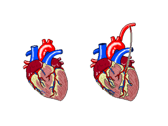
The patient was, until the last year or so, a very fit and active 79y/o, who had suffered from AF for at least 10 years, and had developed a stenosed left anterior descending coronary artery as a result. In addition, echo had shown a severe mitral regurgitation due to valvular prolapse. The procedure would attempt to bypass the LAD using the left internal mammary artery (LIMA), as shown below.
Figure 1: The left internal mammary artery (shown in situ on the right) is relocated from the chest wall to bypass the stenosed section of the LAD.
The plan was to perform the bypass graft, and then set about repairing the damaged mitral valve.
After the patient’s ID and contraindicators had been checked, the anaethetists set about carefully monitoring their respiratory and cardiovascular function as the GA took effect. The ECG, arterial and venous pressure traces were available on lots of screens around the rooms, as whilst they must be monitored all of the time, it becomes particularly important to keep an eye on the given values as the heart is both stopped and re-started.
Interestingly, I noticed a pattern in conversation with each patient throughout the day, as the anaesthetic was administered. The patient was asked to think of their favourite place and the team then asked where that place was. Each time, this was met with silence, but just to make sure, the patient was always asked if they were warm enough. When no answer was forthcoming, they were wheeled into the theatre room proper.
Not everyone on the team was scrubbed up and sterilised, as obviously some would not be required to touch the patient, and others would be required to fetch replacement equipment should it be needed. This created a “sterile field” around the operating table, so only sterile members of staff were permitted within it.
After 1 hour and a whole heap of sterile gowns and drapes were applied over the patient, with only a small window showing the surgical site, the operation began.
Step 1 required access to the thoracic cavity. In case of blood splatter, masks fitted with facial protection were supplied to myself and the other team members who would be in the direct vicinity of the patient when the chest was opened.
An incision was made as illustrated below. This is known as a median sternotomy, and extends from the sternal notch to the xyphoid process. In order to progress past the sternum, an oscillating saw is used to cut throught it. There is a surprising amount of finesse involved in this stage, despite how much pressure is required and as a result, how brutal it appears.
The incision is deepened and cauterised until it travels through the pericardium, so as to allow access to the heart and, after the bleeding vessels around the wound are cauterised, a finochietto retractor is used to hold the sternum open. During this time, the LIMA is found and carefully removed from the chest wall. It is then held in an accessible place with forceps, to be used later.
Lung and cardiac function is transfered to a cardiopulmonary bypass machine, which allows both the heart and lungs to be stopped/emptied, allowing even greater access to the heart due to the lack of lung obstruction, and intricate work to be perfomed whilst the heart is not beating.
The cardiopulmonary bypass apparatus purifies the blood that passes through it from the patient via cannulas placed in the heart and pumps it back, thereby doing the work of the heart and lungs. The heart is fed nutrients at the same time, so as to keep it healthy for the extended period of inactivity. This method of on-pump surgery is known to be incredibly safe- between 1 and 2% of high risk patients will suffer adverse effects as a result of the treatment, and surgical teams are well versed in assessing this via risk factors.
Once the LAD has been correctly identified, an incision is made, creating an opening that roughly matches the size of the end of the LIMA, and the two are stitched together using sutures made of polypropylene,which are no thicker than a human hair, yet can withstand the pressure that would be required to rupture a healthy vessel. To test the suture site, blood is passed through the vessel, and in the event of any gaps in the connection, this blood will be seen outside of the join, which can then be further secured as necessary.
The process, whilst quick to document, was a long one, as each step of the technique was scrutinised carefully before considered complete. In addition, each stitch required at least two people directly, to exact it; one to add the suture, and the rest to support the structures surrounding it.
The second stage of this case was the mitral valve repair, which was in itself a multi-stepped procedure. It is possible to repair a damaged mitral valve using less invasive, keyhole methods, but due to the need for a bypass graft, this wasn’t an option this time.
After gaining access to the valve itself, via an incision in the left atrium, the condition of the valve leaflets is assessed. This particular patient’s posterior leaflet had a prolapsing middle scallop, which meant that the below procedure was necessary to repair it.
The process off attaching the ring was a long one, involving a lot of organisation and intricate knotting. The sutures are applied to both the ring and the valve, the latter is then gently pushed down the threads and into place. These suture strings are then tied off and cut.
Once this has been completed, the valve integrity and functionality was tested by flushing water through the heart. In this case, the valve was still slightly prolapsing, as the water flushed through the valve in an unwanted quantity. The entire process, then, was considered from the beginning, and the valve only said to be repaired, when the regurgitation seen through the valve was minimal.
When performing the annuloplasty removed the surplus anterior leaflet, it took with it a tendineae chordeae, so in order to stabilise it further, an artificial heart string (made of Gore-Tex) was attached to the leaflet and papillary muscle.
A final transesophageal echo was performed, to assess the level of air still present in the heart, and to then assess the function of the repaired mitral valve, and after all of this was successfully accomplished, the long task to gently remove all of the cannulas that were used to bypass the cardiopulmonary system were begun to be removed. Each chamber was sutured in sequence, and the corresponding section of the perfusion apparatus turned off. All of this time, the anaesthetist monitored the patient’s drug infusions and every member of the team monitored pressure and ECG traces. This was still going on whilst the SpR set about cauterising vessels and wiring the sternum before pulling it back together. None of this section of the procedure involved any real finesse; pulling the wires taut so as to close the chest cavity is a test of strength. Each pull caused the patient to move for the first time since coming into the theatre, so I guess that was why it was the only section of the surgery that almost made me wince (something the theatre lead noticed and laughed about). Once it was done, the multiple layers of sutures were applied to seal the surface wound and the patient was taken to recovery.
In all honesty, the most difficult part for me, was in standing for so long. This was echoed by the surgeons, who said that they always felt backed up by their colleagues, so they felt confident that the procedure itself would go well, but standing, often in an unnatural position for so six hours at a time played havoc with their backs and legs. The anaesthetists, being required fully at the beginning and the end of the surgery, spent a great deal of time sat waiting, save for occasionally changing an infusion as and when required. The people working solidly were the surgeons and the scrub nurse, but even the surgeons swapped roles, observed one another without participating, and even took breaks periodically. None of this diminishes any one role, however; each member of the team was required, be it constantly or otherwise, and when one was needed, it took a single word for everyone in the room to know what was required, and by whom. Despite the procedure itself being amazing, it was the mechanical and seamless nature of the professionals in the room that was the most astounding part of the day.
Synap is an upcoming revision tool that is driven by students. The platform enables students to create their own multiple choice questions and upload them, then download those created by others. It’s possible to “follow” other users, as you would someone on Twitter or, incidentally, this site (you can do that in the sidebar of this page…), and take any quiz that they have created. Image upload and basic editing is supported, so quizzes for physiology, such as ECG arrhythmia or echocardiography quizzes are more than possible, and are one of the reasons I decided to get involved with the whole thing. In addition, the app tracks your progress and structures your revision for you, based on your course and modules.
I’ve spent the last week or so beta testing the Synap web platform or, more specifically, I’ve been taking tests and creating basic ECG quizzes to help bug test and check functionality.
The platform, as I’ve mentioned, is currently in closed beta and only present on the web, so without having an app and a larger number of users I cannot comment on it fully, but as it stands, the processes involved in creating a profile and quiz are incredibly simple; adding and annotating images is a cinch, and a complete question only requires the user to add a correct answer and a few wrong ones. Whilst I encountered a few bugs initially, the feedback I provided was swiftly taken on board and the problems were remedied overnight. Taking quizzes is incredibly simple, and all you need to do is click “take quiz” (shockingly), then select your answers and have them marked. You can take these as many times as you like, too, and if the creator has provided any, feedback will be available for each individual question.
My only concern is the reliance on the quiz-maker supplying the correct information. I’ve taken a quiz wherein the correct answer was the only one that was possible to be correct (think “What has tusks and a trunk? 1) Elephant 2)Belephant 3)Your hamster”) yet I was still told my answer was wrong. This is a closed beta, though and that’s what these processes are for. I know it hasn’t escaped the attention of the developers, so we shall see how it is dealt with.
To break all of this down and show you what I know for sure so far, have a look at this (incomplete) features list:
MCQs:
Image/annotation upload
Correct answer & up to 5 incorrect
Optional feedback for test-taker
Optional link to external learning resource
Test result calculation
Obtainable achievements
Personalised revision quizzes sent to you
Community links based on:
Course/Discipline
Cohort
Institution
I’ll add to this list the more familiar I become with the platform.
Omair and James, its creators, and the rest of the Synap team hope that this app will enable students nationwide to help each other and revise together, and it’s a pleasure for me to be involved, even if it’s only in a small way, currently. I’ll continue to post updates as things progress.
(or “what I learned when the tests I have done to others, were done to me”)
The tests presented in this post are intentionally not explained thoroughly here. I have focussed, currently, on patient experience. If you wish to learn more about the things presented here, and the interpretation of the possible results, wait for them to be explained in your lectures, or, perform a quick Google search.
Before the new semester began proper, I was asked to assist with some physiology practicals on my campus. I agreed because I felt (much in the same way a chef will sample his/her food before selling it to the masses) that it would be good for my overall learning to experience the same anxieties and physical exertions -if applicable- that a patient may endure when they undergo physiological testing. When one repeatedly performs tests day in, day out, it’s easy to forget that the patient likely does not have anywhere near the same levels of familiarity with the procedure and proficiency with them as that of you, the practitioner, so to gain an insight into the emotional and physical aspects from the other side would, I felt, be good practice.
Day one was one of a cardio nature, in that I performed lots of exercise tests at a physiologist’s disposal. (Some of these tests are reserved for respiratory physiologists, but if you’re studying and are not yet at the point of choosing which PTP pathway to follow, you’ll experience these, too).
I discovered upon entry, that I would be performing the following:
YMCA step test
Bleep test
Treadmill test: ramp protocol (similar to the Bruce Protocol)
The wait to enter the lab was, (obviously not the same in terms of anxiety levels, but regardless) akin to a patient’s wait to enter a clinic testing room; knowing that I was going to have to perform tests, but not knowing exactly what they were was rather nerve-jangling (especially considering my then-unknown weight gain after the obligatory food-filled, sedentary lifestyle commonly experienced over the festive break).
The real difficulties stemmed from trying to comprehend the techniques required for each test. Explaining, or writing about them is one thing, but actually doing them is another thing entirely.
The YMCA step test itself wasn’t particularly challenging, given that it only involved 3 minutes of steady box steps. The difficulty came in not influencing heart rate on recovery. Knowing that my HR was being documented every minute meant I kept looking at the oximeter, and as has been documented (a quick google search will give you confirmation of this), it is relatively easy to change your HR on command.
For a patient, this last point may not be of particular issue, given that they might not be particularly aware of the potential influence they can have on their HR, but I can easily see how repeatedly stepping onto and off of a box could be difficult task for a patient of advanced age.
Bonus Clinical Perspective: In this test the Heart Rate Recovery and VO2 max doesn’t appear to be particularly accurate, when using normal values, especially when compared with the VO2 max displayed through the other tests, either. The values are based on age, as oppose to individual physiological characteristics, so assume a physical ideal that doesn’t necessarily transcend to real life.
The bleep test wasn’t like ones I have previously attempted in the gym, or what have you; rather, it was more about timing, ensuring there were no stops. This involved slowing down so as to reach the end of the designated track in time with the beep, then speeding up to repeat, meaning that pacing yourself was a must. The resulting strain on my legs caused them to become incredibly painful, incredibly quickly..! (I’m aware that bleep test procedure differs between fitness centres, so forgive my whinging if you use this format regularly).
Encouraging a patient to exhaust themselves doing this test would take a great deal of commitment from both parties; I’m not particularly unfit, but I had nothing tangible to aim for, with regards to an end point, so with no time to “beat”, I didn’t have anything to work towards and as a result, I gave up after 10 or so minutes, despite the fact I could have carried on for a while longer. For the average patient that would frequent clinics to perform this test, achieving maximal exertion may not be something that can be coaxed out of them, especially if they had already endured other tests in the same day.
Already I was beginning to understand the plight of the patient, when it comes to tests that require their full participation, and I still had the hardest one to come… I was not looking forward to the post-lunchbreak activities.
It turns out, the Ramp Protocol test was actually the most enjoyable of the day. Perhaps this was simply because I was growing used to being fatigued/dehydrated, or perhaps it was the setup of the test itself, but I could have happily continued running on the treadmill for a great deal longer than I did, time allowing.
The ramp protocol treadmill test involves the face mask setup presented in the pictures, and a steady speed and incline increase on the treadmill for as long as it takes for the patient to reach their VO2 max, but it is up to the patient when they stop. Unlike the bleep test, which involved travelling at an uncomfortably slow rate at times, the ramp protocol was a fairly rapid journey to a pace similar to that of a distance runner. It was far from comfortable, so would still require a great deal of coaxing and encouragement in order to get the patient to work hard to complete the test, but it was certainly more comfortable than the test that had preceded it.
The whole day not only reminded me of tests and theory that I had almost forgotten, but it really helped me to understand what a patient has to go through when they visit a hospital. The feelings and tests that I personally experienced were, on the whole, not pleasant, but I wanted to be there. For a patient, this will most likely not be the case. When your clinic list is seemingly never-ending and you don’t have time for restarts, it’s easy for the fact that patients don’t know the requirements and procedures as well as you might, to slip your mind, but thanks to this experience, it’s something that I’ll never forget, and I feel it solidifies a vital skill that students require to be able to operate efficiently and fairly: empathy.
Tomorrow brings a different kind of discomfort, in that I will be having my first echocardiogram. I’ll add that experience to part II
The ramp protocol will also get the full write up treatment, as it was by far the most complex and in addition, I have a detailed set of results.
Yesterday, on the 20th of November, Oli and I attended the SCST annual update meeting. It’s the first physiology conference I’ve attended that wasn’t tied to one specific trust (the last one I attended was the Royal United Hospital’s respiratory medicine conference), rather, it was applicable to and attended by cardiac scientists from across the four home nations. The day was packed with talks, networking opportunities and insight into the future of the science. Speakers hailed from a variety of professions and organisations, but all were entrenched in the science of cardiology and education.
Due to the long distance travel and Birmingham’s seemingly city-wide roadworks, Oli and I missed the introduction, but we were present for the rest of the day and we recorded and annotated everything else, so whilst I’ll provide an overview here, detailed breakdowns of everything relevant to PTP study will be supplied separately, as and when time and my coursework volume allows.
Of particular note is the information on preceptorship qualification, delivered by Sophie Blackman of SCST and Boston Scientific. I collared her after the event proper, and she kindly agreed to provide the literature pertaining to this, so as soon as it’s available, I’ll add it for you all to have a mosey over. It seems like a great opportunity for newly- qualified practitioners to become super confident in all aspects of their job, so I highly recommend that you read the contents when they’re available.
Dr Patricia Oakley of King’s College outlined the plans for a new variety of health clinic: the centre that isn’t home and isn’t a hospital, but the “place in the middle”. These will be networked, multidisciplinary centres, featuring social workers, scientists, psychiatrists, GP’s, etc, so cardiac physiologists will most likely be a necessity in their implementation. The whole session really drove home the emerging importance of this profession, but also the requirement of all of us, student and qualified, to ensure that the cardiac physiologist is recognised as being at the forefront of innovation so as not to be overlooked. It was mentioned more than once, that if we don’t put ourselves forward for emerging structures, someone else will.
Dr Oakley told of the need to reduce treatment variability by region. Her example was the treatment of amputation as a result of diabetes; Devon has, by far, the highest number of below-hip amputations when compared with the rest of the UK, due to the fact that the majority of Devonian surgeons trained under a surgeon who has a penchant for this level of removal. The advent of these networked clinics will reduce this level of variability and promote consistency across the home nations.
The president of the AHCS, Dr Brendan Cooper delivered the final talk of the day, discussing the future role of the healthcare scientist in wider healthcare and medicine, and the need for physiologist prescribing. I’ll provide a detailed breakdown of this talk next, and shall hopefully post it in this coming week.
It seems that as soon as I mentioned just how quickly this profession is evolving, something has been raised that enables me to give you an idea of how much.
This blog is called The Student Physiologist. The career’s professionals are known as physiologists or physiological scientists, ergo, myself and my peers are subsequently coined physiology students.
This, however, will soon be a thing of the past, as by the time I qualify, these terms will no longer exist. In their place will be Healthcare Scientist.
It’s difficult to find any sort of identity in such a changing professional environment and this difficulty is bolstered when a physiological scientist tries to explain their role within the NHS. We are among the most patient-facing scientists in the clinical setting, yet we are arguably the least “seen”, in that no matter the description of who you are and what your job is, patients and other staff alike will invariably refer to you as “nurse” or “doctor”. Whilst doctors and consultants are prevalent in this career, it is difficult to convey to patients and staff, the differences between medic and scientist in both the hospital and these roles specifically.
This has highlighted to me, the need for a global identity and perhaps a way for we, as the people with that identity, to forge it for ourselves.
As the evolution moves ever forward, this blog may be named The Healthcare Scientist and I may be signing off with the same name.
We shall see.


 Once this has been completed, the valve integrity and functionality was tested by flushing water through the heart. In this case, the valve was still slightly prolapsing, as the water flushed through the valve in an unwanted quantity. The entire process, then, was considered from the beginning, and the valve only said to be repaired, when the regurgitation seen through the valve was minimal.