Postgraduate Preceptorship

One of the SCST Annual Update sections most relevant to PTP students was based around postgraduate preceptorship. Delivered by Sophie Blackman, of Boston Scientific and the SCST, the talk went through the updates regarding the assessment and accreditation that a newly qualified physiologist can obtain, in conjunction with the council. Sophie stated that she has been quite heavily involved in the production of this framework, and proceeded to outline its intricacies.

The preceptorship programme is relevant to PTP students, because it is aimed specifically at us once we are “let loose”, as it were, into clinical practice. The SCST has received a great deal of feedback from around the country, pertaining to the varying levels at which new healthcare scientists are emerging from their academic study and also feedback from students themselves, on what they feel they need by way of support from the governing body. I for one, can appreciate this; I’m terrified of graduating. I’m confident in my own skills, thus far, and whilst I don’t think that fear will ever go away, I think it’s beneficial for newly qualified HCS to have someone outside of their department that can help them make that transition from student to professional. This nationwide initiative will help to provide this support for the individual, but will also ensure there is an equitable workforce in practice.

The programme itself is based around the individual, and is likely to take anywhere from six months, to two years, based on proficiency. Much like in your current degrees (if you’re a student), the programme features competencies and case based discussions, but in this case, they are undertaken as you perform a job at which you are already doing. Upon “qualification” (this may seem like an odd word to use, given the fact that the practitioner is already qualified, but bear with me…), the student will receive a certificate displaying their confidence in a particular discipline, and that can not only demonstrate a willingness on the practitioner’s part to be the best that they can be, but it will further cement that person’s knowledge and skills base to aid them in their position, thereby helping them, to a point, to leave the student role behind them.

The implication was that if you, as a new HSP, want to make that leap to the STP programme, or follow a different career framework, then this accreditation will assist in identifying your individual fortes, and allow you to perhaps see what pathway you would be best suited to.

The full texts relevant to students are given below:

Preceptorship Framework

Preceptorship guidance for HSPs

For more information, visit the SCST preceptorship page:

new_scst_logo

Screenshot (39)


The Assignment Rush

If you visit this site regularly, or follow the TSP twitter, you’ll hopefully be aware that I try to post updates and tweet fairly regularly. Sometimes they are in the form of personal pieces such as this, sometimes they are reviews or study guides, sometimes minor news updates or small tweaks to the site itself.

If you’ve visited this site recently, however, you might have noticed that I haven’t updated it for a little while. There’s one reason for this, and one reason alone: the assignment.

I’m seeing the light at the end of the tunnel as I type this, due to the fact that my extended case study is now submitted. It was a great piece of work to take on, too. Three sections in total, each with its own word count. The first was a patient case study, with a provided patient history, two electrocardiography traces to analyse and compare, and the subsequent report and treatment pathway to complete. This was probably the most fun of the three, but also the most time consuming.

The second was a broad topic that had to be condensed into a relatively small word count. This was, without question, the most difficult of the three sections and whilst I’m confident in the content I submitted, it’ll be interesting to read my feedback and see how well I interpreted the question.

The third, a brief similar to the second section only with a much more specific topic was initially fairly free-flowing, in terms of writing, but the further I progressed through it, the further away that word count looked. What began as a torrent of information on the page, soon trickled to a halt, as I realised I had explored all of the avenues in my plan without leaving room for branching out. As a result, I had to go back through every paragraph and find some wiggle room to potentially add another facet to the discussion.

I’m being intentionally vague so as not to disclose any details of the assignment, rather I’m confirming what we all know about assignments; there are some we like, and there are some that we loathe.

If you’re currently in the midst of a large piece of work, don’t panic! Just power through. Don’t worry about your friends posting Facebook updates from the pub, don’t worry about your Christmas shopping just yet. I know it’s easier said than done, of course, but despite the fact that (technology permitting) this article has gone live at around 3pm, as I type this, it’s 4.30am. Pressing that ‘submit’ button on my coursework has sent me into a state of fatigued euphoria, and all the late nights have been worth it.

I’ll add that on top of this assignment, I also have deadlines for an Inter-professional Collaboration self-reflective essay and a presentation on cardiac technology in the same week. Next week.

Until then, it’ll be a bit quieter around here…

Screenshot (39)

Review: simECG: ECG Simulator v1.186

Download for Windows/Linux: Free

Developers: Antonio Cardoso Martins, Paulo Dias Costa, Joao Miguel Marques


I’ve been searching for a half-decent ECG simulator since last year, but hadn’t found one that costs less than “more than I have”, so I was pleasantly surprised to find the rather unnecessarily named simECG: ECG Simulator for free, on Windows and Linux.

simECG offers a number of functions in its current version. The operator can select from a series of common arrhythmias at the click of a button, and observe the associated waveform on the display. Unfortunately, only a handful of options are actually selectable, at present, with the others showing as greyed out, presumably, as with many Open Source programs, until they are finalised by the development team.

simECG screen 2 (2)

The custom settings tab provides the means to alter each area of the trace individually, adjusting heart rate, P wave amplitude/duration and more, and watching the displayed trace change in real time. The program hints at future save/load functions for your altered settings, too, which will be a nice inclusion for educators to make use of.

simECG screen 1

All of the aforementioned are easy to use and clearly marked, even if there aren’t currently all that many of them.

The option exists to change the background between ECG paper and a monitor screen, although the ECG paper skin is purely cosmetic. It would have been nice if the paper option was more in correlation with the amplitudes and durations selectable in the readout options. Greyed out sections of the “preferences” tab hint that calibration will soon be able to be changed by the user, so it would be preferable for beginners and students if these proposed calibration options had a realistic background to use in conjunction with the created trace.

simECG screen 4

I couldn’t find an option to reset the trace at all, even in a greyed out form, and as a result, returning to the default custom settings is something of a chore. Hopefully this is something the developers will consider including in future iterations.

By now you may have noticed the appearance of the waveforms in the above trace. The trace waveform was one of the first things I noticed, as the whole thing doesn’t look right. The P and T waves look malformed, with the latter presenting almost as though the patient was displaying hyperkalaemia despite this being labeled as a normal sinus ECG.

simECG screen 3

The assessment quiz tab gives the user an opportunity to identify 10 rhythms in 60 seconds. It’s fun, sure, but given the odd appearance of the waveforms, it becomes a case of memorising the traces present in this program alone, as they aren’t all applicable to real life.

I’ll be honest, it’s hard to criticise something that the developers admit will “never be finished” due to its Open Source status, but the nature of this website and Open Source in general means it pays to remain objective. In actuality, whilst I have highlighted a few issues, the fact that this tool is ever-evolving and totally free, means I can only commend the development team for their ethos and hard work.

Martins, Costa and Marquez state their belief that education shouldn’t be a corporate tool, or purchasable commodity, rather it should be accessible to all. The more people there are to flag issues, the better an idea the team can have of what functionality to add, what bugs to fix, and what other changes are felt to be necessary by users. Despite being generally incomplete at present, it’s not only one to watch for in the future, but one I’d ask every cardiac physiologist to download and play around with.

Due to this version still being in the 1.n phase, I have high hopes for the future of this software, as it has great potential as a learning tool. With the addition of more options in the preset tab, further wave/interval customisation, and more accurate waveforms in general, simECG could help physiology students consolidate their knowledge without carting loads of textbooks around, making it an essential bit of kit.

Screenshot (184).png


Screenshot (39)

Review: Analyze ECG Reporting

Download for Google Play: Free

Download for iOS: Free

Developer: Cathal Breen

Analyse phone


When you’re just beginning to get to grips with analysing a 12-lead, taking a methodical approach is recommended, but in practice that’s easier said than done. Remembering what you’re measuring, and in what order you’re measuring it, is sometimes confusing, especially when, like me, you’re still getting your head around the various concepts behind the plethora of arrhythmias and pathological morphologies you’re likely to find in a patient ECG.

Screenshot_2015-11-21-17-09-26~2

I’ve already covered the tutorial apps documenting normal and abnormal values that I felt were most beneficial to PTP students, but Analyze ECG Reporting by Cathal Breen exists solely to guide the practitioner through each, single analysis and serves as a methodical reminder of everything that should be documented in your report.

Analyse is nicely presented, with a very simple user interface set up for each section. The display contains boxes for measured values, buttons to advance to the next measurement, or to go back to make corrections and some pop-up menus for comments on the ECG waves. It doesn’t suffer from a text overload, or clutter in any way. The colour scheme is visually appealing, but conservative, so when using the app, you’re kept on task and not distracted by needless images or too many different colours.

Screenshot_2015-11-21-17-35-28~2

This app is merely a way to educate practitioners into using the same approach to study each trace. It may seem like an obvious thing to point out, as ECG cannot provide a diagnosis on its own, but Analyse is not an algorithm that will diagnose a pathology for you.
This is not to diminish this app’s merits whatsoever, though. Analyse does a great job of clarifying the process of ECG analysis and provides a list of the necessary things to include when reporting. Since installing it, I have used it to methodically review lots of the traces I’ve obtained, including those set in my coursework.

In fact, my only problem with Analyse ECG Reporting is in correcting mistakes from the drop down menu. A long press on the option that you have selected will remove it from the final list, but this isn’t explained at any point. It took me a little while to figure it out, so some brief instructions wouldn’t have gone amiss upon starting up the app for the first time. It’s a minor niggle and it didn’t detract from my overall experience with Analyse, however.

Screenshot_2015-11-21-17-09-51~3

Consistency and a methodological approach are key parts of analysis, and Analyse ECG Reporting is a great trainer. This app is a must have for PTP students, but I’d recommend it to any student who’ll have more than a passing dalliance with electrocardiograms.

Screenshot (180)


Screenshot (39)


Interview With A Distance Learner

The fact that this specialist degree exists primarily in universities is a relatively new event; before the shakeup by Modernising Scientific Careers, the majority of training was completed in-house with an element of distance learning thrown in to assist with the theory behind the practical concepts.

As physiological science makes the transition to a 100% university- led discipline, there remain students that are still learning the “old way”. Sarah is one of those people, and I had the pleasure of working with her this year during my rotations between respiratory medicine and cardiology. In order to get a bit of insight into exactly how the course differs between bases, she kindly agreed to be interviewed for TSP.

Hux-Ham

Hello Sarah! Could you outline the structure of your week, with regards to working in your department and studying the degree simultaneously?

I’m employed by the hospital, so have to work my set hours which are Monday – Friday 08.30-16.30. Although I’m studying, I am not employed as a student, rather, I am an Assistant Technical Officer, which basically means I help around the department doing admin, portering and some clinical work. I have certain responsibilities with regards to admin that I have to keep on top of regardless of what clinical work I need to be learning.

Monday is my main admin day, so I spend the entire day sorting through referrals, checking messages & booking appointments for certain procedures that only I book. I need to keep on top of this as some of the procedures have extremely long waiting lists, so if a patient cancels last minute I need to try my best to fill that slot. Once my admin is complete I normally help out my colleague in the office with some of her work load. If there is no porter to bring inpatients up & back for echocardiograms then it is part of my job to do this as well, which means I can’t get my necessary admin work completed.

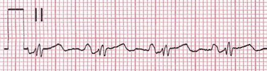
Tuesday is the start of my clinical week, unless I have been portering the previous day. At the moment I am spending all day Tuesday in analysis, analysing 24 hour and 48 hour tapes. I am able to analyse a tape independently, but as I am still learning they all need to be checked after, just in case I’ve missed something or worded my report incorrectly.

Wednesday is a half day in the department for me as I have a collaborate session starting at 12.00 so I need to be set up in the library ready to start. After my collaborate session I catch up on any studying I need to do, such as looking over lectures that have been released for the following week, researching/ writing an assignment or revising for upcoming exams. On a Wednesday morning I will either be fitting ambulatory blood pressure monitors (supervised, as I am not confident to do them alone yet) or analysing.

Thursday mornings I am in Electrocardiography, either in the department or going down to the ward, and in the afternoon I analyse.

Friday mornings I do tape clinic which occupies the entire morning and keeps me very busy, especially if I have patients returning that have had symptoms of dizziness & I need to get the tapes checked before I can let them go. I spend Friday afternoons in analysis.

That is my current working week, but I will start going on the rota soon to sit in on exercise treadmill tests as well. Most mornings I get into work at around 07.30 so I can get some studying done before work and I try to do an hour or so in the evening as well. Most weekends I keep to myself, but if I have an assignment due or exams I will do a couple of hours each day.

That’s a hectic week. This might now be a silly question, but do you feel that this is this enough?

In terms of clinical exposure … yes! But it is very hard to keep up with the academic work load when there is very little time to fit things in. I commute for over 2 hours a day so this eats into my potential study time, but I try to keep a balance of work, study and actually having a life!

Do you feel that working in the same department as you study helps you to learn more and keep you motivated?

I feel that second year especially has helped me learn, but most of the academic work in our first year wasn’t particularly relevant to cardiology. I feel like I learnt more in the last 2 months from analysing tapes than I have in the whole 2 years that I’ve worked in the department. I definitely think it has helped to keep me motivated as I’m constantly surrounded by people that are doing the job I am training for, so I’ve got a clear goal at the end of it.

You’re one of the last sets of the distance intake. Do you think, if you had the choice, you’d still do the degree in the manner you currently are, or would you choose to be based at the university?

I’ve already done a previous degree so I’ve experienced the whole student life thing, so I’m not missing out by doing it this way. At the moment I am essentially being paid to learn, which is ideal. I wouldn’t be able to afford to do this degree if I was based at the university, as I’ve already had a student loan so I’m not entitled to another. I think I get a good amount of exposure in the clinical setting, but I just have to do some of the boring admin jobs to make up for it. At the end of my degree I will have a job and I know 100% that this is the career I want for myself. I wasn’t passionate about my previous degree subject so I lost interest and didn’t want to spend the rest of my life doing it, whereas I know from working in this department and from studying the way I am, that this is what I want to do. I don’t think I’d have that level of clarity if I was based more at the university than the hospital.

That’s fair. When we worked together during my placement, I was aware of the fact that you were much more comfortable in the clinic environment than I was (obviously), so what do you feel we at the university have by way of an advantage?

I definitely think that as I’m exposed to patients and the environment all day every day that I am more confident and comfortable than yourself, but I would say that full time students based at the university have a lot more academic knowledge. We have 1/2 hours a week of contact time with our lecturers so we need to go out and research ourselves, whereas it is clear that you guys have a lot more academic time although you miss out a lot with the lack of placement.

Thanks, Sarah!

As you can probably tell, despite the fact that Sarah and myself are in the same cohort, our academic years have a vastly different focus. As I (rightly) assumed just from working with her on the department, both routes present their pros and cons, and seeing as this is a vastly understaffed form of diagnostic science, it does, in my opinion, open the career up to a greater number of people now it will be university- led.

If you’ve got an opinion, or a question regarding anything you’ve read, sound off in the comments below.

Photo courtesy of Facebook

Screenshot (39)

I Have LVH, Should I Be Worried? Normal Variants Of An ECG

As part of our course we often perform ECG’s on one another to enhance and refine our practical ECG skills. During a practical recently I volunteered to be the patient so that my fellow students could practice their electrode placement skills; whilst being filmed and critiqued by others. The group universally agreed in the value of this experience, despite the fact it felt strange performing an ECG to an audience of your friends whilst being filmed in an artificial clinical environment. After everyone had practiced we printed of a recording of my ECG. Our lecturer, whom is a senior physiologist, explained that we would as a class analyse the ECG in our following lecture.

Upon the analysis of my ECG, my lecturer broke the news to me that I had left ventricular hypertrophy (LVH) with sinus bradycardia. Considering the previous lectures we’d had on LVH and its clinical significance I was pretty scared few a seconds or so. He then thankfully reassured me that in my case this was completely normal. My lecturer emphasised the importance of always combining your ECG analysis and findings within the context of your patient.

My ECG LVH sinus brady

He explained my athletic physique (his words) and my age were enough to convince him that my development of LVH was not due to pathological reasons but that of heart remodeling as a result of prolonged physical conditioning. If presented with a 30 stone, 60 year old male, with LVH, or a 70 year old sinus bradycardic female ticking a long at 50 bpm and suffers occasional syncope; this would not fit as a normal finding in the context of these patients.

This practical highlighted the importance of always putting your ECG findings in the context of your patient and the normal ECG variants that may be encountered. The patients we see will be varied. They will be of different genders, ethnicities, ages, physical condition, possibly even pregnant. All these groups will produce significant normal variants in their ECG’s. These factors must always be taken into consideration when assessing the significance of your findings. Is your patient bradycardic and symptomatic suffering frequent dizzy spells or are they a young physically conditioned adult. We will be posting articles shortly on the normal variants expected in some of these patient demographics.

What is LVH

LVH is an increase in size and proportion of ventricular myocardium (in this case specifically the left ventricular myocardium). This can occur in any chamber of the heart but is most commonly found in the LV.

How is LVH diagnosed on an ECG

Most trained operators will spot signs of LVH relatively quickly on a ECG recording as it will be common that precordial ventricular tracings will overlap one another requiring a reduction in gain settings.

However, the Sokolow Lyon criteria is correct method of choice. This involves measuring the amplitude of the S wave in V1 and adding it to the R wave amplitude in V5 OR 6 (whichever bigger). If the sum amplitude is greater than 3.5mV LVH is suspected.

To calculate right ventricular hypertrophy (RVH): Amplitude of R wave in V1 + S wave V5 or 6. If the sum amplitude is greater than or equal to 1.1mV RVH is suspected.

However, an ECG alone cannot determine the extent of hypertrophy and its clinical consequence. An echo-cardiogram would be required to ascertain this.

Causes of LVH

LVH is a result of increased demand put on the LV to increase cardiac output. Over prolonged periods of time this increases cardiomyocyte size. As discussed earlier, this increased CO demand could be the result of exercise, and therefore sustained activity levels could lead to LVH. The upshot of this is that for every heart contraction the ventricles can force out a greater volume of blood for every beat (stroke vol) reducing the demand on the heart to supply cardiac output at rest. For this reason athletes with LVH will have a lower resting HR (sinus bradycardia).

However, there are also many pathological reasons for developing LVH, all of which result in pressure overload in the LV increasing its resting workload.

Some of the most common pathologies associated with LVH are as follows:

• Hypertension (most common cause)

• Aortic stenosis

• Aortic regurgitation

• Mitral regurgitation

• Coarctation of the aorta

• Hypertrophic cardiomyopathy

As LV hypertrophy develops, the myocardium can become so thickened that it begins to inhibit the filling of the LV reducing cardiac output leading to increased risk of mortality.

In my next article I will be looking further into some of the pathological causes of LVH and their clinical significance.

Ref:

http://lifeinthefastlane.com/ecg-library/basics/left-ventricular-hypertrophy/

The Lewis Lead

Scouring the web for cheap textbooks, I happened upon something of which I wasn’t even remotely aware: alternate ECG lead placements. As has been highlighted in previous posts by myself and others, cardiac scientists have strict guidelines that ensure we perform an ECG procedure to an accurate and repeatable standard, so it came as a bit of a surprise to discover that there existed a different way of carrying out the test with an aim to view specific activity.

The Lewis lead, named after Sir Thomas Lewis, is an alternate placement that can be used to better view atrial activity in relation to that of the ventricles. In many ECGs, it can be rather difficult to assess P waves; whilst they are represented using standard lead configuration, they are much less apparent than ventricular activity, due to the nature of the ECG’s detection mechanisms.

Using the Lewis lead configuration, it is possible to increase the detection of atrial activity and diminish that of the ventricles and gain a clearer picture of atrial fibrillation, flutter and, in the case of the article that brought my attention to this system, improving P wave recognition in wide QRS complex tachycardia.

The configuration is as follows:

Lewis Lead

  • RA electrode on the manubrium
  • LA electrode on the 5th IC space, right sternal border
  • LL on the right lower coastal margin
  • RL remains in the standard SCST position
  • Adjust calibration to 20mm/mV

As shown on the diagram, a three lead configuration is still present, as in Einthoven’s triangle, but Lead I now travels directly over atrial activity. For this reason, Lead I is used as the monitor lead and the one from which a rhythm strip should be taken.

On the trace itself, there is a marked visual difference. The following were recorder on the same patient and we begin with the standard electrode configuration:

normal-lead-I-II

And now introducing the Lewis lead setup:

s5-atrial-lead-i

s5-atrial-leads-iiThe P waves present in these altered leads are much more pronounced.

There are more lead systems that are used in the diagnosis a variety of different conditions such as Brugada syndrome. I’ll research and cover these and try to get some more traces using the Lewis lead system throughout the year.

Traces courtesy of ekgcasestudies.com

Screenshot (39)

How Is Your ECG Electrode Placement?

As a student cardiac physiologist it has been drilled into our heads from an early stage the importance of correct anatomical electrode placement in obtaining an accurate ECG recording. An ECG measures the electrical activity of a patient’s heart from many different angles, and is achieved by placing 10 sticky electrodes on the patient; four on the limbs and six on the chest. For correct electrode placement we follow the clinical body guidelines set out by the our governing body, the SCST. As specialists within the field, we have a duty to perform these tests in a standardised, methodical manner to produce reliable and accurate diagnostic information, as the ECG is the first port of call when assessing heart abnormality.

Screenshot (17)

Unfortunately, from my experience, and from that of my colleagues, the misplacement of these electrodes has become somewhat commonplace. To the unassuming operator this may seem superficial but incorrect placement of electrodes can alter the ECG patterns displayed simulating or concealing abnormalities, such as myocardial ischemia/infarction.

There is evidence that many health professionals who record ECG’s have not been suitably trained or assessed in the technique: A study by Kings College London into electrode misplacement highlighted that only 50% of nurses and less than 20% of cardiologists correctly place leads V1 and V2 during a standard 12-lead ECG. These numbers are quite shocking and highlight the widespread misunderstanding of this key diagnostic tool.

Close0up
An example of how NOT to perform an ECG. V1 and 2 are incorrectly placed, as are 3 and 5.

I personally witnessed an example of this whilst on my first week of placement. I was performing an ECG on a patient within the cardiac ward under the supervision of an assistant technical officer who regularly performs ECGs. I correctly located the anatomical landmarks on the patient’s chest and applied the electrodes, as per the official guidelines. At this point, the ATO interrupted me and challenged the placement of my V1+V2 electrodes, stating they were too low. She then took over control of the procedure and removed the electrodes. She began to count the intercostal spaces, beginning from the clavicle. The guidelines state the operator should identify the manubriosternal joint, or angle of Louis, on the patient to locate the second intercostal space as their first anatomical landmark. This subsequently meant her V1 and V2 electrodes were placed too high and  my original placement was in fact correct. After the procedure I challenged my colleague about this explaining we were taught to follow the SCST guidelines in our electrode placement. The ATO responded by saying that this was “how they had always done it.” I discussed this with my clinical educator and the issue was later addressed with my colleague.

The consequence of incorrect ECG recording can lead to potentially incorrect diagnoses and inappropriate treatment leading to wasteful use of healthcare resources and even cause harm to patients. Evidence suggests that adequate training of operators reduces ECG recording errors. However as the SCST highlights in their guidelines, the indications there is little awareness in many practitioners of the need for training.

Clearly, the solution to this issue is to increase awareness in health professionals exposed to ECG practice about the importance of correct electrode placement.  This could be achieved by increased collaboration between cardiac physiologists and other healthcare professionals. As specialists within the field we have duty to share our expertise and knowledge to ensure our patients receive the best standard of care. As a profession we should be much more active in teaching and increasing awareness of what we do and why it is so important. Relevant staff should be confident in performing ECGs not because of experience, but due to high quality training and continual auditing.

To achieve this I feel our profession needs to embrace this responsibility and be far more active in the support and training of other health professionals.

Khunti, K. (2013) Accurate interpretation of the 12-lead ECG electrode placement: A systematic review. Health education journal . 73 (5) pp. 610-623.

Harrigan, H., Chan, TC., Brady, JW. (2012) Electrocardiographic Electrode Misplacement, Misconnection, and Artifact. The Journal of Emergency Medicine [online]. 43 (6), pp. 1038–1044.

Baxter, S, Blackman, S, Breen, C, Brown, C, Campbell, B, Cox, C, Eldridge, J, Hutchinso, J, Rees, E, Richley, D, Ross, C. Society for Cardiological Science and Technology (2014) Recording a standard 12-lead electrocardiogram. Available from: http://www.scst.org.uk/resources/CAC_SCST_Recording_a_12-lead_ECG_final_version_2014_CS2v2.0.pdf

http://millhillavecommand.blogspot.co.uk/2012_05_01_archive.html

Screenshot (39)

Have You Ever Tested A Robot? Pt II

I still haven’t.

Bear with me, though, as this is going somewhere, I swear.

After the last session, in which I provided the robot’s voice and controlled its HR and ECG, it dawned on me that as a result, everyone had the opportunity to be filmed performing the test and gain valuable group feedback, except me.

I wasn’t the only one to notice this, as it transpired.

During a subsequent lab session, wherein we practiced manual BP, honed bedside manner, discussed contraindications and compared different methods of BP measurement, it was revealed that the remainder of our ECG feedback period would be completed in the lab. We no longer had immediate access to the simulation mannequin, so thanks to a willing volunteer, another of my colleagues was able to complete the procedure and again receive feedback in a partitioned area of the lab.

Then it was my turn to step up to the plate.

I was the last to ‘go’, as it were. The difference between my assessment and the other’s lies in that everyone else enjoyed an element of seclusion: the curtains around the bed-space being pulled in the first session and the high walls that separated one section of the lab from the other, in the second. The rest of the group stayed outside of these boundaries in everyone else’s case. Not for me, though. I stood away from the couch, preparing to make my entrance to the imaginary treatment room I could see in front of me and just before I could open the invisible door, the consultant physiologist taking the session said “Wait, I’m just going to call everyone else in, if that’s ok?”

“…If that’s ok”, as if I had a choice.

Everyone else filed in. They kept filing in for what felt like an age. My lecturer, the rest of my class and the head of physiology. Then, they all looked at me, waiting.

I’m not sure how I’d have fared if I’d known this was going to be the format for my peer assessment, but I feel no shame in admitting that I don’t remember ever being as scared as I was before I started moving. I didn’t know how to begin, so I just went for it. I walked into the ‘room’ (after, somewhat embarrassingly, opening the invisible door) and performed the test as I would out on placement.

I asked all the required questions and added one or two patient identifiers to account for the fact that I didn’t call my patient from any waiting room and gained a consented, accurate trace.

Not only did I do it all with the eyes of more than a couple of people scrutinising my every move, I did it with a piece of equipment I have never used before and the most tentacle-like cable configuration I’ve ever seen in my life- if you’ve tried to untangle the wires behind your television when you’re moving house, you’ll know what I mean but, trust me, this was worse. In addition, I managed to ignore a completely new experience: the fact that I was so scared that the back of my neck was sweating..!

Fear is natural. It’s normal to be scared of doing something that’s relatively new to you, especially when you know you’ll be watched and judged doing it. Whatever ‘it’ is, it wouldn’t feel like a real achievement if we didn’t feel fear beforehand. I’m glad it was sprung on me, if I’m honest. My final assessments and various practical examinations for the rest of my career will follow this format so it’s good to have a grasp on some of the emotions I’ll be feeling before them. If you’re just beginning the PTP programme, you’ve got things like this to look forward to, so just try to enjoy it. Realise that the fear of these things is normal and, most importantly, the sooner you take a deep breath and swallow the lump in your throat, the sooner they’ll be over!

Thanks!

Screenshot (39)

Light At The End Of The Tunnel

Having being around for a few years now, I’ve read an ECG or two in my time.  If you’re still early on the road to becoming a fully-fledged Physiologist though, let me assure you of one thing:  IT DOES GET EASIER!

I won’t lie, even with the experience I now have, there are still the occasional strips that leave me scratching my head like a confused monkey but on the whole, a 12-lead doesn’t scare me anymore.  One thing that I think many students will find at some point during their learning, is that their more experienced counterparts have somehow forgotten how difficult it is to read an ECG. You might take an ECG for someone to check and receive a reply along the lines of ‘Well, obviously this is…’ Not all that helpful!

Learning to read an ECG is a lot like learning to read a new language. Sure, if you’ve been practicing for a long time, you’re pretty fluent, but it’s important to remember how hard you found it back when it was still just a foreign language to you. Only then can you start to empathise with those who are in that position now. And if you are in that position now, don’t give up!

If I could offer one word of advice to you, the person reading this who is desperately trying to get to grips with ECG, it would be this: get to know what a normal ECG looks like really, really, REALLY well. Then, get to know how that relates to the electrical and mechanical activity of the heart. (I suppose that’s sort of 2 pieces of advice but stick with me here). If you can get all of that into your head, you’re putting yourself the best possible position for progression.  If you instantly know what a normal ECG looks like, any abnormality should stand out like a sore thumb. You might not know what the abnormality is, but if you know how the ECG waveforms relate to the mechanical activity of the heart, you can at very least a take good, educated guess on what that abnormality suggests the heart is actually doing. You won’t be an expert, not at first, but you will have the foundations on which you can build and make yourself one.

Therefore, the most important first step is to learn what is ‘normal’ but I’ll discuss that in detail in a later post.